Biomedical Engineering Reference
In-Depth Information
8.1 Bacterial Biofi lm Infections on Implant Materials
A biofi lm is an aggregate of bacteria in which bacterial cells adhere to each
other on a wet or moist surface. Biofi lms may form on living or non-living
surfaces and can be prevalent in natural, industrial and hospital settings
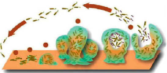
[1, 2]. The formation of a biofi lm (as shown in Figure 8.1) begins with the
attachment of free-fl oating bacterial cells to the surface. As the bacterial
cells propagate quickly, the biofi lm structure develops becoming more
complicated. At the last stage, the bacterial cells release into the environ-
ment and contaminate other surfaces.
Biofi lms are considered easy to form but hard to treat, which can cause
wide-spread infections [4] in the human body, for example, through cath-
eter infections, infections on the inert surfaces of artifi cial implants [5] and
the formation of dental plaques [6] (Figure 8.2). Statistics show that bio-
fi lms are involved in an estimated 80% of all infections [7]. These biofi lm
infections can be serious and hard to treat because the development of the
biofi lm structure may allow for bacteria to be increasingly antibiotic resis-
tant, because the bacteria in the biofi lm are held together and protected by
a matrix of EPS (extracellular polymeric substance or exopolysaccharide).
This matrix protects bacteria cells within it and facilitates communica-
tion among them through biochemical signals, resulting in their increased
resistance to detergents and antibiotics. In some cases the effect of anti-
biotic resistance can be increased a thousand-fold [8]. Thus, bacteria in
biofi lms are frequently related with persistent infections [9] in the body.
An implanted medical device provides a surface for bacteria to attach
to and multiply in a patient's body, resulting in the formation of a biofi lm.
Bacterial biofi lm infection is a problem for all implants. For example, infec-
tion is one of the most common causes for failure of a hip implant, respon-
sible for 14% of the total number of revision surgeries [11]. Another example
4
5
3
2
1
Figure 8.1
The biofi lm life cycle [3]. 1: individual cells populate the surface.
2: EPS (exopolysaccharide) is produced and attachment becomes irreversible.
3 & 4: biofi lm architecture develops and matures. 5: single cells are released from
the biofi lm.
Search WWH ::

Custom Search