Biomedical Engineering Reference
In-Depth Information
7.2.1
Architecture of Bacterial Cell
Prokaryotic cells have several distinct surface layers that are briefl y men-
tioned below.
Capsules
: If the high molecular weight polysaccharides fi lm deposits
strongly on the cell wall, it is called a capsule, and otherwise, it is known
as an extracellular material slime. The main function of these layers is to
avoid phagocytosis [28].
Cell wall
: Gram staining classifi es bacteria on the basis of different
cell wall structures. A class of bacteria that are stained by gram stain are
named as gram positive bacteria and the rest are known as gram nega-
tive bacteria. The gram strain reacts with the peptidoglycane layer of bac-
teria and produces a crystal violet color [29]. The peptidoglycane layer
is comparatively thick and peripheral to the cell wall in gram
−
positive
bacteria. Whereas, this layer is very thin in gram
negative bacteria and is
located inbetween the inner membrane and outer membrane [30, 31]. The
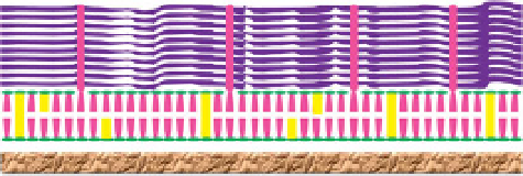
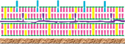
difference in cell wall of both gram positive and gram negative bacteria is
shown in Figure 7.5. Other important constituents of the cell wall include
the following:
Peptidoglycan
: Peptidoglycane is a long strand of alternating polymer
of
N
−
−
acetylmuramic acid (NAM) and
N
−
acetylglucosamine (NAG) [32].
Highly
−
thick and crosslinked peptidoglycan layer is present in gram
−
positive cells, while it is very thin in gram
negative cells. The peptidogly-
can layer is the main target of antimicrobial activity [33].
−
LPS
Lipoprote
Peptidoglycan
Outer membrane
Periplasmic space
Phospholipid
Inner membrane
Protein
Cytoplasm
(
a
)
LTA
Peptidoglycan
Phospholipid
Cytoplasmic
membrane
Protein
Cytoplasm
(
b
)
Figure 7.5
Cell wall structure of (a) gram
−
negative, and (b) gram
−
positive
bacteria (adapted from ref. [23]).


Search WWH ::

Custom Search