Biomedical Engineering Reference
In-Depth Information
(
a
) Tooth
(
b
) Pulp-dentin organ
Dental
pulp
Dentinal tubules
Odontoblastic
processes
ECM-sensing
cytoskeleton
Integrins
500 nm
Pulp ECM
Hydroxyapatite-
crystals
(
c
) Dentin
nanostructure
(
b
) Odontoblast interaction
with ECM
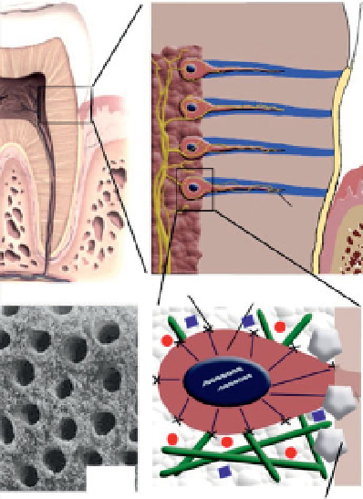
Figure 6.9
Macro-nano structure: Dentin/pulp organ. (a) Illustration of tooth
at organ level (macro level). (b) Odontoblast cell position in the pulp/dentin
(micro level). (c) Enlarged illustration of the odontoblast interaction with pulpal
non-mineralized and mineralized extracellular matrix (nano level). (d) Dentin
micro/nano structure may help in the reconstruction of pulp-dentin organ
scaffold.
“Braille-read” its bed through the environment-sensing cytoskeleton and
integrin receptors (Figure 6.9). The information carried from ECM by
actin-myosin forces propagates to the cytoskeleton-caged nucleus initiat-
ing intracellular signaling cascades that ultimately alters genes expression
to direct the cellular tissue-specifi c spatiotemporal behavior (adhesion,
contraction, migration, proliferation, differentiation, self-renewal and
apoptosis) [104]. Macroscopically, dentin is a calcifi ed tissue forming the
stress-bearing body of the tooth and bearing odontoblastic processes.
However the properties and mechanics of the environment around the
odontoblast cell might be very different from the macroscopic properties
of the tissue they weave. An odontoblast in contact with the dental pulp
soft matrix and the dentin hard surface provides an example of a hier-
archically structured microenvironment where mechanically and biologi-
cally different matrices impact the cellular behavior.
Search WWH ::

Custom Search