Biomedical Engineering Reference
In-Depth Information
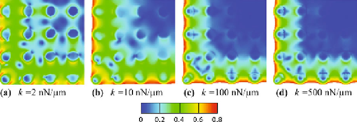
Fig. 3.4
Contour plots of the distribution of stress fiber orientations, characterized by the quan-
tity
Γ
, for a cell in steady state adhered to a post-bed with post stiffness (
a
)
k
2nN
/
µm,
(
b
)
k
=
10 nN
/
µm, (
c
)
k
=
100 nN
/
µm, and (
d
)
k
=
500 nN
/
µm. Only quarter segments of the
square cells are shown
=
higher focal adhesion densities near the cell periphery and the polarized horseshoe
shape on individual posts. The fact that there is a significant density of contrac-
tile stress fibers is reflected in the curvature of the cell edge visible in Fig.
3.3
(b).
The heterogeneous nature of the cell response is more pronounced in this case than
for the more compliant posts, evidenced by the degree of alignment of stress fibers
shown in Fig.
3.4
(b). The distribution of stress fiber orientations is almost uniform
at the center of the cell, whereas they are highly aligned along the cell edge. This
and other features visible in Figs.
3.3
(b) and
3.4
(b) indicate that posts near the cell
perimeter are interacting with other perimeter posts to a significant degree via the
stress fibers, and are interacting with the cell as a whole. Furthermore, the stress at
the center of the cell is relatively high, only falling rapidly near the cell edges in a
shear lag phenomenon. The stress gradient there is what gives rise to the horseshoe
shape of the focal adhesions on posts near the cell perimeter, as the force applied
by the cell to an individual post has a net resultant acting towards the open side of
the horseshoe. A high, almost uniform stress at the cell center explains the relative
lack of focal adhesions on posts near the cell center (Fig.
3.3
(b)), as the absence of
a stress gradient means that little force is being applied to the post tops in that area.
Next, we consider the simulations for posts with even higher stiffness,
k
=
10 nN
/
µm, with the focal adhesion distribution shown in Fig.
3.3
(c). In this case,
spatial gradients of stress near the cell perimeter are steeper than for more compli-
ant posts. As a result, the concentration of focal adhesions on posts near the cell
periphery is even higher than for the more compliant posts, and the horseshoe shape
of the focal adhesion distribution on these posts is more distinct. Note also that
the concentration of focal adhesions on posts near the center of the cell is lower
than for the more compliant posts, indicating a more uniform stress there when the
posts are stiff. The contractile stress and the concentration of stress fibers in the cell
are greater than for the more compliant posts, as can be deduced from the higher
curvature of the cell edge visible in Fig.
3.3
(c). The heterogeneous nature of the
response of the cell is apparent in the degree of alignment of the stress fibers, shown
in Fig.
3.4
(c), with an almost uniform distribution of orientations in a large patch at