Biomedical Engineering Reference
In-Depth Information
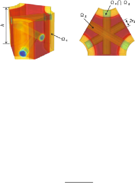
Fig. 2.5
Illustration of the molecular-level unit cell. The collagen fibrils are connected by the
GAGs via next-nearest neighbor connectivity. The subdomains are:
Ω
c
Ω
g
—the overlapping do-
main of the coating and interconnecting GAGs;
Ω
g
—the region occupied only by interconnecting
GAGs;
Ω
c
—the region occupied only by the coating GAGs
∩
where
e
is the unit charge supplied by the disaccharide unit. The uniform charge
density for the coating GAGs is
λ)
ρ
eff
Ah
V
c
ρ
c
=
(
1
−
,
(2.26)
where
h
is the length and
Ah
the volume of the unit cell, respectively. The bridg-
ing GAGs repeat along the axis of collagen fibrils with period
h
, which may be
determined using the conservation of total charge
N
gag
i
1
l
i
g
αe
b
=
ρ
eff
Ahλ
=
,
(2.27)
where
λ
is the charge fraction for the bridging GAGs and
N
gag
i
1
l
i
g
is the total length
of the GAG rods over the unit cell. As an example, we employ a next-nearest neigh-
bor topology for the interconnecting GAGs proposed by Muller et al. (
2004
). The
3-D unit cell model has uniform charge density
ρ
c
in the coating domain and
ρ
g
in
the bridging GAG cylinders (Fig.
2.5
).
The Poisson-Boltzmann equation is solved for the electrostatic potential
ϕ
over
the cell subdomains with boundary conditions as described by Eqs. (
2.21
) and (
2.22
)
and with fixed charge density
ρ
f
prescribed as,
=
⎧
⎨
ρ
c
on
Ω
c
,
ρ
f
=
(2.28)
ρ
g
on
Ω
g
,
⎩
ρ
c
+
ρ
g
on
Ω
c
∩
Ω
g
,
and
ρ
f
=
0 elsewhere. Charge conservation is applied to the bridging and coatings
GAGs domains independently. As the unit cell is deformed, the charge density
ρ
g
changes due to the cylinder length change. The coating charge density
ρ
c
will not
change, as discussed above. For simplicity, the GAG radius
r
g
is invariant during
cell deformation.