Biomedical Engineering Reference
In-Depth Information
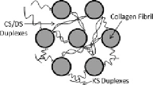
Fig. 2.1
Collagen-PG
arrangement in the corneal
stroma; some of the GAG
chains may bridge fibrils by
antiparallel duplexing (Scott,
1992
)
2.1 Introduction
The extracellular matrix of the cornea is composed of two principal molecular com-
ponents: type I collagen in the form of 25 nm diameter fibrils and small leucine-rich
repeat proteoglycans (PGs). The corneal stroma is organized into approximately 500
lamellae (or fibers) through its thickness and within each lamella the collagen fib-
rils are maintained in almost perfect parallel arrays with a quasi-regular hexagonal
packing arrangement. The collagen is responsible for carrying the tensile forces that
are produced by the intraocular pressure. The corneal PGs consist of linear chains
of disaccharide units covalently bound to a core protein. Predominant corneal gly-
cosaminoglycan (GAG) components are dermatan sulfate (DS), chondroitin sulfate
(CS) and keratan sulfate (KS). Scott (
1992
) proposed that some GAGs form inter-
fibrillar bridges by duplexing and this has been supported by some evidence from
imaging (Muller et al.,
2004
; Lewis et al.,
2010
). These arrangements are illustrated
in Fig.
2.1
. The DS, CS and KS disaccharide units are ionized at physiological
pH and carry two negative charges per unit. The electrostatic interaction of these
charges with ionic species gives rise to strong intermolecular forces that are respon-
sible for the tissue osmotic pressure.
The transparency of the cornea requires that the collagen fibrils be maintained in
their lattice-like arrangement. Modeling the forces of interaction between the colla-
gen fibrils and GAGs may provide insights into the mechanisms underlying corneal
transparency and is a primary goal of this work. The polyelectrolyte nature of the
corneal stroma is well illustrated by its remarkable capacity for swelling when im-
mersed in water or dilute salt solution. The tendency of the corneal stroma to swell
can be characterized by the equilibrium swelling pressure. The equilibrium swelling
pressure may be measured by compressing a piece of isolated corneal stroma in an
ionic bath solution between permeable plates until equilibrium is reached (Hedbys
and Dohlman,
1963
). During the past several decades, the swelling pressure on var-
ious species have been measured experimentally (Hedbys and Dohlman,
1963
; Fatt,
1968
; Olsen and Sperling,
1987
), and it has been observed that the swelling pressure
is highly dependent on the tissue hydration.
Several previous investigations have aimed to create theoretical models for
corneal swelling (Hart and Farrell,
1971
; Hodson,
1971
; Olsen and Sperling,
1987
).
It has been demonstrated that Donnan theory for osmotic pressure is incapable of
fully explaining swelling pressure (Olsen and Sperling,
1987
). In this work we pro-
pose a swelling pressure theory that is derived from a molecular-level description of