Biomedical Engineering Reference
In-Depth Information
Table 3.2 Optimal coil
orientation and angle of the
precentral gyrus with respect
to the interhemispheric cleft
at cortical hot-spot position
for each subject
Subject
Optimal coil rotation
Angle of gyrus
220
46
:
5
Ch
200
38
:
5
Fe
210
39
:
5
Ha
210
30
Pa
230
54
Ro
210
39
St
43
:
75
As subject 'Ti' only participated in session 1, we use the mini-
mum of that session
40
Ti
3.1.3 Relevance for TMS
The monotonicity in our measurements documents that we have identified reliable
minima with our setup. The recordings show that there is no valid single optimal
coil orientation for stimulation of the foot for all subjects. The standard lateral coil
orientation, however, is not optimal. The optimal coil rotation for stimulating the
right foot (abductor hallucis muscle) deviates approximately 30
from the standard
coil orientation. The MT difference of optimal coil rotation to standard rotation
was of 11
:
8 and 10
:
8 % of MSO, respectively (Fig.
3.5
).
Furthermore, our recordings support the assumption of a sinusoidal relationship
between coil orientation and stimulation outcome—in this case with the motor
threshold as quantitative parameter. The result of the fitting (cf. Fig.
3.4
) however
mainly relies on the recordings around the minima. Due to our setup, no recordings
for the maxima region exist. With more recordings in the maxima regions the
sinusoidal curve may slightly change. However, the general trend—due to the
minima—should remain. Note that this model also fits well to the data presented
by Balslev et al. for the hand region [
4
].
(a)
(b)
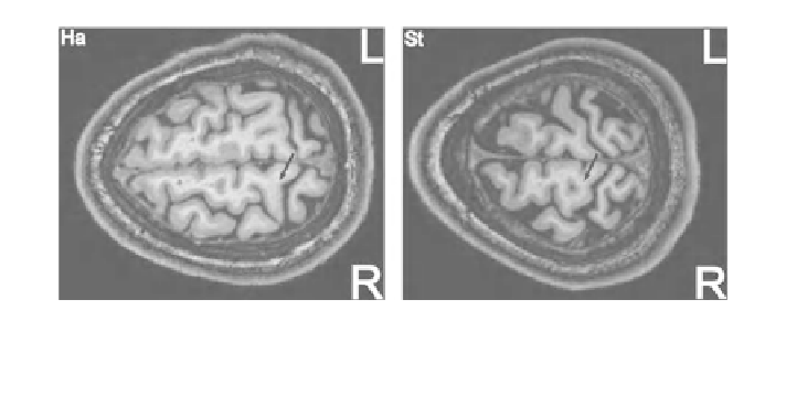
Fig. 3.5 Hot-spot (starting point of the black arrow) for two subjects projected in the MRI
images in a transversal view. An area of the precentral gyrus at the edge to the central sulcus and
close to the interhemispheric cleft is in focus for stimulation. The black arrows denote the found
optimal coil orientation for the individual subject


Search WWH ::

Custom Search