Biomedical Engineering Reference
In-Depth Information
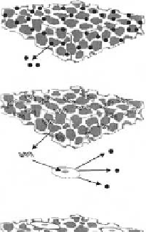
(a)
Fertilized ovum
Embryonic stem (ES) cells
(b)
Somatic (adult) stem cells
Blood cells Kidney
CNS
PNS
Skin
Heart
Differentiated cells
(c)
Fig. 7.2-28 From undifferentiated cells to differentiated cells.
hierarchy structure as represented in
Fig. 7.2-28
. The
starting cell for human body development is a fertilized
ovum, which then forms an embryo as a result of re-
peated cleavages. The modura formed after 3-4 day and
the blastocyst formed after 5-7 day cleavage contains
pluripotent stem cells, named ''embryonic stem'' (ES)
cells. Circulating blood cells survive for only a short time
ranging from days to months. Throughout the entire life
they are replenished by hematopoietic stem cells in bone
marrow which provide a continuous source of pro-
genitors for red cells, platelets, monocytes, granulocytes,
and lymphocytes. In addition to hematopoietic stem
cells, adult bone marrow contains also non-hematopoietic
stem cells. The stem cells for non-hematopoietic tissues
are referred to either as MSCs, because of their ability to
differentiate into cells that can be roughly defined as
mesenchymal, or as bone marrow stromal cells (BMSCs),
because they appear to arise from the complex array of
supporting stromal structures found in marrow. It has
often been stated that a key factor in tissue engineering is
lineage-committed precursor cells, especially multi-
lineage stem cells, but almost differentiated cells also
have large applications in the current tissue engineering.
Many different types of stem cell exist, but they all
are found in very small populations in the human body; in
some cases 1 stem cell in 100,000 cells in circulating
blood. To identify these rare types of cells found in many
different cells and tissues, scientists use stem cell
markers. Each cell type has a certain combination of re-
ceptors on their surface that makes them distinguishable
from other kinds of cells. In many cases, a combination of
multiple markers is used to identify a particular stem cell
type.
Table 7.2-9
lists some of the markers commonly
used to identify stem cells and to characterize differen-
tiated cell types.
Fig. 7.2-27 Three primary polymeric growth factor delivery
strategies: (a) growth factors are embedded within the polymer
matrix and released; (b) genes encoding a growth factor are
embedded within the polymer matrix and released, followed by
cellular uptake and expression of the gene to produce growth
factor; (c) growth factor is released from cells seeded on the
polymer matrix that secrete the factor.
tissue formation and repair, important variables in for-
mulations for delivery systems include the concentration,
timing, and sequence in which the growth factors are
introduced.
To circumvent the difficulty of sustained release, high
cost, and low commercial availability of growth factors,
several methods have been attempted for the sustained
delivery of growth factors. As shown in
Fig. 7.2-27 [25]
,
the methods used to deliver growth factor molecules in-
clude the development of systems to deliver the protein
itself, genes encoding the growth factor, or cells secreting
the growth factor. Injection of recombinant plasmids and
transplantation of gene-manipulated cells have been
performed with an expectation that growth factors will be
continuously produced from the modified cells for a certain
period of time. The DNA most widely employed for such
studies is those which are responsible for the biosynthesis
of VEGF that induces vascularization. Plasmid vectors are
relatively safe but vulnerabletonucleaseattackandcon-
sequent inefficiency and expense. Viral vectors including
adenoviruses, retroviruses, lentoviruses, etc., increase
gene transfer efficiency but themselves have limitations
including possible toxicologic and immunologic re-
sponses. Retroviruses are expressed only in proliferating
cells and permanently integrate into genomic DNA.
7.2.8 Cell sources
7.2.8.1 Differentiated cells
A human body consists of approximately 60 trillion cells.
Human cells differentiate from stem cells into pro-
genitor, precursor, and mature cells, forming a tree-like
Previous thinking was that many differentiated cells of
the adult human have a limited capacity to divide, but