Biomedical Engineering Reference
In-Depth Information
view of the effect of pseudo-craniofacial development on
the subject. Thus, developmental visualization is essen-
tially a 3D morphing technique that uses biologically ap-
propriate control functions to describe the morphing.
Also, it is important to notice that these morphing tech-
niques attempt to morph (i.e., translate, rotate, scale)
every voxel in the volume, unlike the more conventional
morphing techniques used for 3D surfaces. Volumetric
morphing is computationally intensive; however, it can
avoid topological problems, such as self-intersection, that
3D surface morphing can easily encounter.
modality, mostly on the same subject (
Figs 6.6-5 and
6.6-6
). Such spatial comparisons or localizations help to
validate the efficacy of examinations. Often, direct cor-
relation measures do not provide a good quantitative
description of the correspondence between the data
because statistical variations deteriorate the correlation
measure. Multimodality volumetric visualization pro-
vides correlation information and helps the user to vi-
sualize the presence of spatial localization in the data.
When the two sets of data have different spatial orien-
tations, automated 3D registration may be difficult and
interactive visualization may help explore and un-
derstand the data. The technical challenges in this class
of volume rendering are not demanding, but the logic of
combining two or more volumes requires attention. The
data may need to be combined pixel by pixel, or pixels
may need to be substituted from image to image. The
Multimodality visualization
3D visualization can be used for morphological correla-
tion between different forms of volumetric data, such as
those acquired using different types of imaging exams or
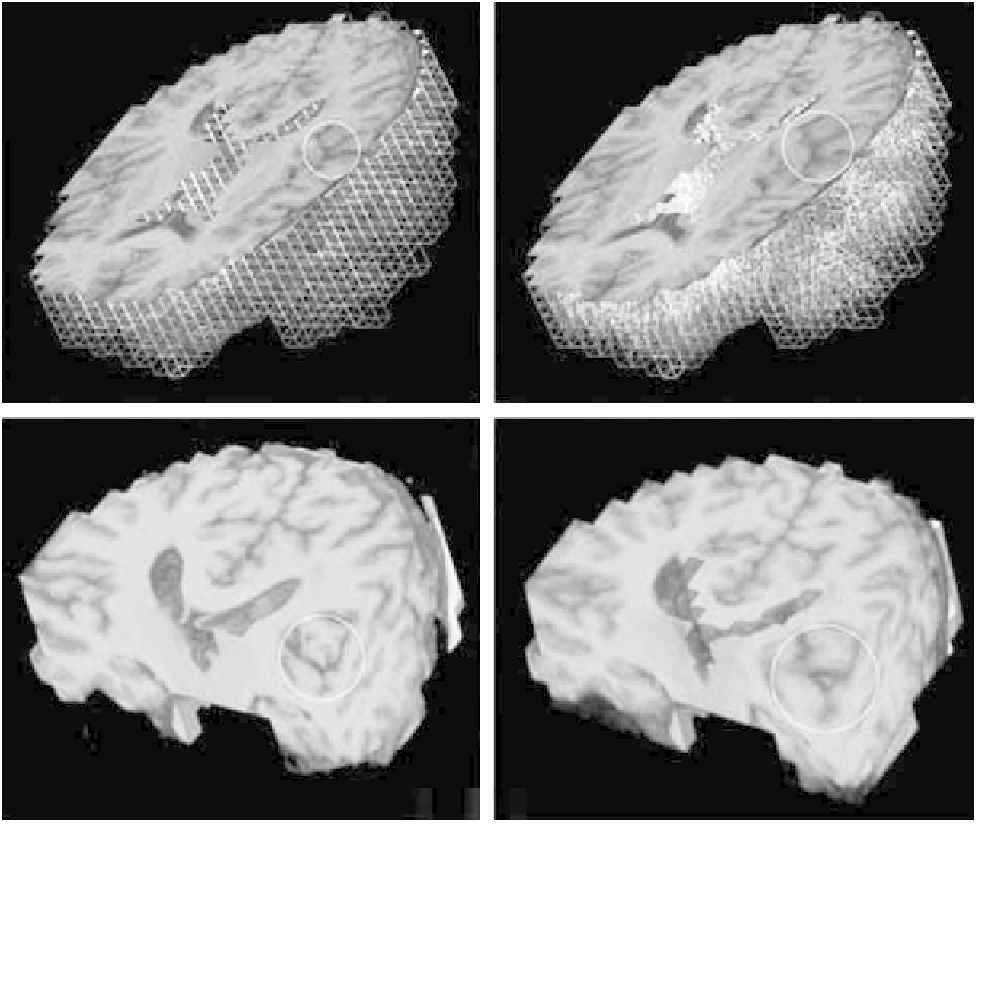
Figure 6.6-12 Finite-element modeling of tumor growth and its visualization using 3D morphing to visualize the morphological changes
in the surrounding anatomy. (Top) FEM mesh (white: white matter, gray: gray matter, red: boundary nodes). (Bottom) FEM embedded
3D visualization. (Left) Undeformed brain. (Right) Deformed brain. (Images courtesy of Stelios Kyriacou, M. Solaiyappan, Christos
Davatzikos.)