Biomedical Engineering Reference
In-Depth Information
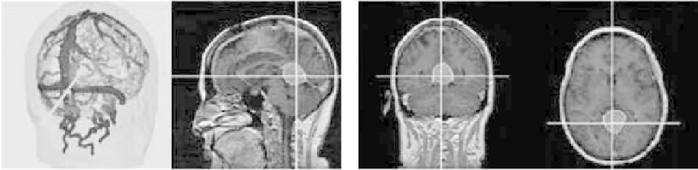
Figure 6.5-7 Pointer tracking in 3D MRI rendering and three orthogonal MRI slices.
6.5.2.4 Visualization subsystem
movements of the LED configuration attached to
the head clamp.
(6)
Proceed with craniotomy and surgical procedure.
(7)
At any point, use sterile Flashpoint pointer to
explore structures in the MR imagery.
Two types of visualizations are provided to the surgeon on
the workstation monitor. One is an enhanced reality vi-
sualization in which internal structures are overlaid on
the video image of the patient. The video image is set up
to duplicate the surgeon's view of the patient. Any seg-
mented MR structures may be displayed at varying colors
and opacities (see
Fig. 6.5-5
).
A second visualization shows the location of the
pointer tip in a 3D rendering of selected MRI structures
and in three orthogonal MRI slices (see
Fig. 6.5-7
). These
visualizations are updated twice per second as the pointer
is moved.
6.5.4 Performance analysis
To evaluate the performance of our registration and
tracking subsystems, we have performed an extensive
set of controlled perturbation studies
[10]
. In these
studies, we have taken existing data sets, simulated
data acquisition from the surface of the data, added
noise to the simulated surface data, then perturbed the
position of data and solved for the optimal registration.
Since we know the starting point of the data, we can
measure the accuracy with which the two data sets are
reregistered.
Although extensive details of the testing are reported
in Ettinger
et al.
[10]
, the main conclusions of the anal-
ysis are as follows:
Accurate and stable registration is achieved for up to
45
rotational offsets of the data sets, with other
perturbations.
Accurate and stable registration is achieved for up to
75
rotational offsets of the data sets, with no other
perturbations.
Robust registration is obtained when the surface data
spans at least 40% of the full range of the surface, and
is generally obtained with as little as 25% coverage.
Small numbers of outliers do not affect the
registration process.
6.5.3 Operating room procedure
Using our system, as seen from the surgeon's perspective,
involves the following steps:
(1)
Prepare patient for surgery as per usual procedure,
including clamping the head. Head is still visible.
(2)
Attach a configuration of LEDs to the head clamp,
and record the positions of the LEDs in the
Flashpoint system.
(3)
Register MRI to patient by placing our scanner bar
over patient's head. The bar is generally about 1.5 m
away from head. Scan patient's head by swabbing
a trackable probe across the skin. Typically several
swabs are used, designed to cover a wide range of
positions on the patient. It is often convenient to
include swabs along known paths such as across the
cheeks or down the nose, as these paths will aid in
inspecting the resulting registration.
(4)
The Flashpoint/laser bar may be repositioned at any
point to avoid interference with equipment and to
maintain visibility of LEDs.
(5)
Sterilize and drape patient. Any motion of the
patient during this process will be recorded by
6.5.5 Operating room results
We have used the described image-guided neurosurgery
system on more than 100 patients. These cases included