Biomedical Engineering Reference
In-Depth Information
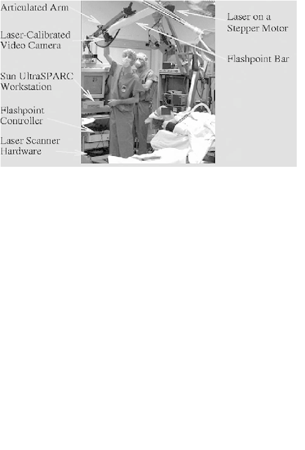
Figure 6.5-2 Physical setup for image guided surgery system.
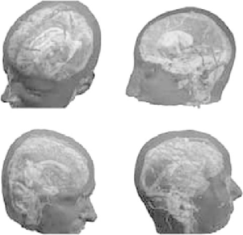
The result is an augmented, patient-specific, geo-
metric model of relevant structural and functional in-
formation. Examples are shown in
Fig. 6.5-3
.
[12]
, benefiting from its dense data representation, but
use either a laser scanner to construct the patient's scalp
surface or a trackable probe to obtain data points from
the patient's skin surface for registration.
We have used two related methods to register the
reconstructed model to the actual patient position. In the
6.5.2.2 Registration subsystem
Registration is the process by which the MRI or CT data
is transformed to the coordinate frame of the patient.
Excellent reviews of registration methods include
[22,
24, 32]
.
Extrinsic forms of registration use fiducials (e.g.,
[1,
21, 31, 35]
): either markers attached to the skin or bone
prior to imaging or anatomically salient features on the
head. The fiducials are manually localized in both the MR
or CT imagery and on the patient, and the resulting
correspondences are used to solve for the registration.
Fiducial systems may not be as accurate as frame-based
methodsdPeters
et al
.
[28]
reports fiducial accuracy
about an order of magnitude worse than frame-based
methods, but Maciunas
et al
.
[21]
reports high accuracy
achieved with novel implantable fiducials.
Intrinsic registration is often based on surface align-
ment, in which the skin surface extracted from the MRI
data is aligned with the patient's scalp surface in the
operating room. Ryan
et al
.
[29]
generates the patient's
scalp surface by probing about 150 points with a track-
able medical instrument. Colchester
et al
.
[6]
uses an
active stereo system to construct the scalp surface. We
also perform the registration using surface alignment
Figure 6.5-3 Examples of patient reconstructions by segmenting
MRI scans into different tissue types.