Biomedical Engineering Reference
In-Depth Information
strengths of three techniques: single-channel expecta-
tion/maximization segmentation, binary mathematical
morphology, and active contours models. Masutani
et al.
[73]
segment cerebral blood vessels on MRA images
using a model-based region growing, controlled by mor-
phological information of local shape.
Many segmentation techniques developed originally
for two-dimensional images can be extended to three
dimensionsdfor example, region growing, edge de-
tection, or multispectral segmentation
[12, 19, 21, 90,
125]
.
3D segmentation
combined with 3D rendering
allows for more comprehensive and detailed analysis of
image structures than is possible in a spatially limited
single-image study. A number of 3D segmentation tech-
niques can be found in the literature, such as 3D con-
nectivity algorithm with morphological interpolation
[57]
, 3D matching of deformable models
[70]
, 3D edge
detection
[78]
, coupled surfaces propagation using level
set methods
[131]
, and a hybrid algorithm based on
thresholding, morphological operators, and connected
component labeling
[46, 100]
.
There has been great interest in building digital volu-
metric models (3D atlases) that can be used as tem-
plates, mostly for the MR segmentation of the human
brain
[23, 47, 61]
.A
model-based segmentation
is
achieved by using atlas information to guide segmenta-
tion algorithms. In the first step, a linear registration is
determined for global alignment of the atlas with the
image data. The linear registration establishes corre-
sponding regions and accounts for translation, rotation
and scale differences. Next, a nonlinear transform (such
as elastic warping,
[5]
) is applied to maximize the simi-
larity of these regions.
Warfield
et al.
[122, 123]
developed a new, adaptive,
template-moderated, spatially varying, statistical classifi-
cation algorithm. The algorithm iterates between a classi-
fication step to identify tissues and an elastic matching step
to align a template of normal anatomy with the classified
tissues. Statistical classification based upon image in-
tensities has often been used to segmentmajor tissue types.
Elastic registration can generate a segmentation by
matching an anatomical atlas to a patient scan. These two
segmentation approaches are often complementary.
Adaptive, templatemoderated, spatially varying, statistical
classification integrates these approaches, avoidingmany of
the disadvantages of each technique alone, while exploiting
the combination. The algorithm was applied to several
segmentation problems, such as quantification of normal
anatomy (MR images of brain and knee cartilage) and pa-
thology of various types (multiple sclerosis, brain tumors,
and damaged knee cartilage). In each case, the new algo-
rithm provided a better segmentation than statistical
classification or elastic matching alone.
Figure 6.4-11
shows an example of 3D segmentation
of normal and pathological brain tissues. The tumor
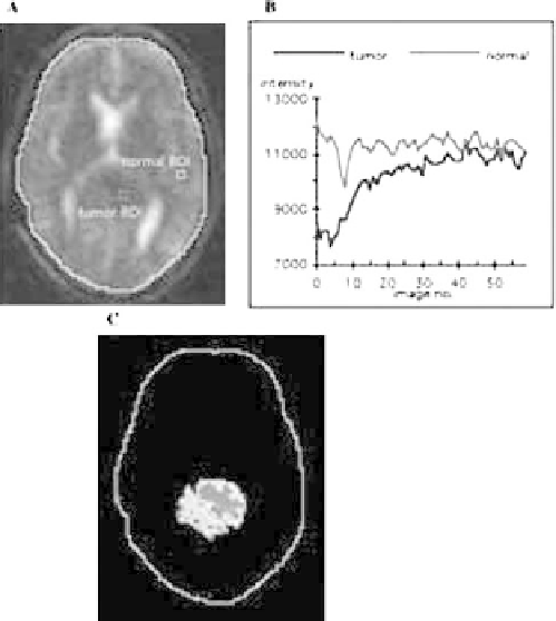
Figure 6.4-10 Image segmentation using correlation mapping.
(A) First image in a sequence of 60 temporal images with 3 3
pixel ROIs drawn in tumor and normal area; (B) plot of the average
intensity of the reference ROI (tumor) and the normal ROI for 60
images in a sequence; (C) correlation map of the tumor.
interest in the tumor area and a normal ROI is shown in
Fig. 6.4-10
A.
Figure 6.4-10
B plots the average intensities
of the reference and normal ROIs. The correlation map is
displayed with a pseudocolor lookup table in
Fig. 6.4-10
C.
The technique of correlation mapping has found
numerous applications. Some of them are included in
Refs
[92, 93]
. Other investigators have adopted this
technique in brain activation studies
[7]
, segmentation of
breast tumors
[71]
, and renal pathologies
[108]
.
A modification of correlation mapping technique,
called
delay mapping
, is also used to segment temporal
sequences of images. It segments an image into regions
with different time lags, which are calculated with re-
spect to the reference
[94]
.
Parametric maps, similarity maps, and delay mapsd
all are segmentation and visualization tools for temporal
sequences of images. They are particularly useful for
evaluation of disease processes, drug treatments, or radi-
otheraphy results.
6.4.7 Other techniques
Combined
(
hybrid
)
strategies
have also been used in
many applications. Here are some examples: Kapur
et al.
[58]
present a method for segmentation of brain tissue
from magnetic resonance images that combines the