Biomedical Engineering Reference
In-Depth Information
It is between these two radii that the ray associated with
the assumed plane-wave solution is constrained to move.
Outside these two values of
r,
called caustics, the ray
becomes imaginary, leading to decaying fields.
For a fixed value of b,as
y
increases, the region be-
tween the two caustics becomes narrower. As
y
is in-
creased further a point will be reached where the caustics
merge. Beyond this point the wave is no longer bound.
The propagation conditions of a wave depend on the
value of both b and y. For a fixed value of y, modes far
from cutoff have large b values and correspondingly more
closely spaced caustics. In general, a bound hybrid mode
in a graded-index optical fiber can be represented pic-
torially by a skew ray spiraling down the fiber between
two caustics. Both inside (
r < r
1
) and outside (
r < r
2
) the
caustics, the field corresponding to the hybrid mode
decays.
transmitted undisturbed to the receptor. The scattered
x rays merely interfere with the information conveyed by
the shadow pattern of transmitted rays. Therefore,
a mechanical grid is inserted behind the patient to pre-
vent most of the scattered x rays from reaching the
cassette. For a parallel, monoenergetic x-ray beam in-
cident along the
z
axis, the distribution
N
(
x, y
)of
transmitted x-ray photons at the image plane is given, in
the absence of scattering, by
N
0
A
ð
e
m
ðzÞ
dz
where the line integral is taken over all tissues along the
unscattered photon trajectory to the point (
x, y
) on the
image plane, m is the linear attenuation coefficient for
x rays of the tissue encountered at (
x, y, z
), and
A
is the
x-ray energy absorption coefficient of the intensifying
screen. The distribution of x rays absorbed in the screen
thus forms a two-dimensional projection image of the
transmission of x rays through the three-dimensional
volume of tissue exposed to the x-ray beam.
The linear attenuation coefficient m is in fact the sum
of the coefficients for various types of x-ray interactions.
For the range of x-ray energies employed in medical
imaging, two kinds of interactions predominate: the
photoelectric effect, described by the linear attenuation
coefficient s, and Compton scattering described by
the linear attenuation coefficient s. Thus m
¼
s
þ
s
.
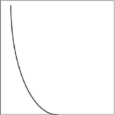
Figure 6.2-19
shows the x-ray energy dependencies of
these coefficients in human soft tissue. The photoelectric
coefficient increases with atomic number
Z
like
Z
3
,
principally because x rays interact photoelectrically with
6.2.5 CT scanners in medicine
For almost a century, x rays have been used for medical
imaging and radiation therapy. Over 100 years ago
Wilhelm R¨ntgen, a professor of physics at the Julius
Maximilian University of Wurzburg, discovered x rays
while experimenting with cathode rays in a Crookes
tube. Word of this discovery spread quickly, and by early
1986 the properties of x rays were under investigation in
many laboratories in Europe and North America. By the
turn of the century, physicians were exploiting the pen-
etrating character of x rays to look inside the human body
without cutting it open.
The usage of x rays for medical diagnosis and therapy
has expanded enormously since those early years. Today,
in the United States alone, over 300 million clinical x-ray
examinations are performed annually for purposes rang-
ing from static imaging of fractures and cancers to the
real-time guidance of tissue biopsies and cardiovascular
angioplasties. In addition, half a million cancer patients
each year receive x-ray treatments, about half of them for
curative purposes and the rest for pain relief.
Until recently the diagnostic and therapeutic appli-
cations were distinct. Today, however, the boundary be-
tween the diagnostic and therapeutic applications of
x rays in medicine is far less distinct.
Ordinary planar x-ray images are formed by placing
a patient between an x-ray tube and an image receptor,
usually a cassette containing an intensifying screen and
a photographic film. The film is exposed by light emitted
when the transmitted x rays interact in the screen. The
resulting radiograph is a static shadow image. Fluoros-
copy is a variant of this procedure in which a fluorescent
screen and an electronic image intensifier are used to
form a continuous moving picture. When the x rays
traverse the patient, they can be absorbed, scattered or
1.0
Compton
0.10
Photoelectric
0.010
0
50
100
150
PHOTON ENERGY (keV)
Figure 6.2-19 Linear attenuation coefficient for x rays traversing
human soft tissue is the sum of two dominating contributions.