Biomedical Engineering Reference
In-Depth Information
category are neuropathies (from diabetes), nerve lesions,
depressions and anxiety. The latter category may include
emotional disorders and lie detection. Qiao et al. (1987)
developed a method to measure skin potential, skin
electrical admittance, skin blood flow and skin temper-
ature simultaneously at the same site of human palmar
skin. This was done in order to be able to investigate
a broader spectrum of responses to the activity of the
efferent sympathetic nerve endings in palmar blood
vessels and sweat glands.
Both
evoked
responses (e.g. to light, sound, questions,
taking a deep breath) and
spontaneous
activity may be of
interest. Measurement of spontaneous EDR is used in
areas such as sleep research, the detection of the depth of
anesthesia and in sudden infant death syndrome research.
Figure 4.1-27
shows detection of EDR by means of
88 Hz conductance (
G
) and susceptance (
B
) measure-
ments (Martinsen et al., 1997a). The measurements
were conducted on palmar sites in right and left hand. In
Fig. 4.1-27
both hands show clear conductance waves,
but no substantial susceptance waves. No time delay can
be seen between the onset of the conductance waves in
the two hands, but the almost undetectable susceptance
waves appear a few seconds after the changes in con-
ductance. This indicates that these changes have differ-
ent causes. The rapid conductance change is presumably
a sweat duct effect and the slower change in susceptance
is most probably due to a resultant increased hydration of
the stratum corneum itself. There are no susceptance
waves that could indicate any significant capacitance in
the sweat ducts.
Venables and Christie (1980) give a detailed sugges-
tion on the analysis of EDR conductance waves based on
the calculation of amplitude,
−
20
0.85
Conductance
0.8
−
16
0.75
−
12
0.7
0.65
−
8
0.6
Potential
−
4
0.55
0
0.5
0
50
100
150
Time (s)
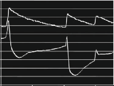
Figure 4.1-26 Simultaneous registration of exosomatic AC
conductance and endosomatic DC voltage. Source: Courtesy of
Azar Jabbari.
and polarization basically appear in parallel in biological
tissue.
The
endosomatic
measurements are carried out as DC
voltage measurements. The mechanisms behind the
changes in skin potential during symphaticus activity are
not known, but processes like sodium reabsorption across
the duct walls and streaming potentials in the sweat
ducts should be taken into account.
Figure 4.1-26
shows
that the origin of the endosomatic and exosomatic curves
is not identical.
The so-called ''lie detector'' is perhaps the most well-
known instrument in which the electrical detection of
this activity is utilized. There are, however, several other
applications for such measurements, mainly within
the two categories; neurological diseases or psycho-
physiological measurements. Examples of
the first
latency, rise time and
18
16
G
right
14
12
G
left
10
B
right
8
6
B
left
4
2
0
0
10
20
30
40
50
60
70
80
Time (s)
Figure 4.1-27 Measured 88 Hz admittance GSR activity on palmar skin sites. A deep breath at approximately 20 seconds on the
time scale triggered the response.