Biomedical Engineering Reference
In-Depth Information
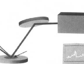
Monochromator/
Photmultiplier
1300 to
4000 cm
−
1
532 nm
SFG
Sample
OPG/OPA
Nd: YAG Laser
1064 nm
Fig. 3.1.4-18 Schematic diagram of a SFG apparatus (based upon a diagram developed by Polymer Technology Group, Inc.).
spectroscopic technique has been severely limited for
surface studies because of its low signal level. However,
in recent years, great strides in detector sensitivity have
allowed Raman to be applied for studying the minute
mass of material at a surface. Also, surface enhanced
Raman spectroscopy (SERS), Raman spectra taken from
molecules on a roughened metal surface, can enhance
Raman signal intensity by 10
6
or more. Raman spectra
will be valuable for biomedical surface studies because
water, which absorbs radiation very strongly in the in-
frared range, has little effect on Raman spectra that are
often acquired with visible light (
Storey
et al.
, 1995
).
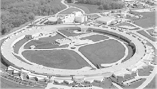
Synchrotron sources of energetic radiation that can be
usedtoprobematterwereoriginallyconfinedtothephysics
community for fundamental studies. However, there are
nowmore synchrotron sources, better instrumentation, and
improved data interpretation. Synchrotron sources are
typically national facilities costing
>
$100M and often oc-
cupying hundreds of acres (
Fig. 3.1.4-19
). By accelerating
electrons to near the speed of light in a large, circular ring,
energies covering a broad swath of the electromagnetic
spectrum (IR to energetic X-rays) are emitted. A synchro-
tron source (and ancillary equipment) permits a desired
energy of the probe beam to be ''dialed in'' or scanned
through a frequency range. Other advantages include high
source intensity (bright light) and polarized light. Some of
the experimental methods that can be performed with great
success at synchrotron sources include crystallography,
scattering, spectroscopy, microimaging, and nanofabrication.
Specific surface spectroscopic methods include scanning
photoemission microscopy (SPEM, 100 nm spatial resolu-
tion), ultraESCA (100
m
m spatial resolution, high energy
resolution), and near edge X-ray absorption spectrometry
(NEXAFS).
Studies with surface methods
Hundreds of studies have appeared in the literature in
which surface methods have been used to enhance the
understanding of biomaterial systems. A few studies that
demonstrate the power of surface analytical methods for
biomaterials science are briefly described here.
Platelet consumption and surface
composition
Using a baboon arteriovenous shunt model of platelet
interaction with surfaces, a first-order rate constant of
reaction of platelets with a series of polyurethanes was
measured. This rate constant, the platelet consumption
by the material, correlated in an inverse linear fashion
with the fraction of hydrocarbon-type groups in the
ESCA C1s spectra of the polyurethanes (
Hanson
et al.
,
Fig. 3.1.4-19 The Advanced Photon Source, Argonne National
Laboratories, a modern synchrotron source.