Biomedical Engineering Reference
In-Depth Information
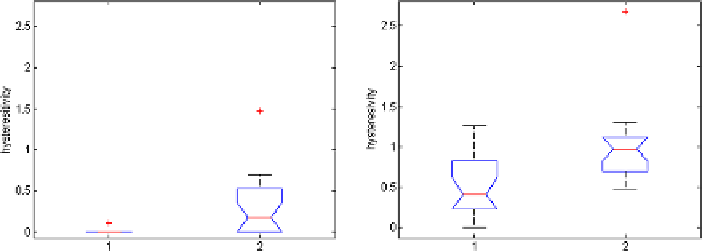
Fig. 7.5
Tissue hysteresivity
η
r
with FO2,
p<
0
.
0012 (
left
) and with FO4,
p<
0
.
0004 (
right
);
1
: Healthy subjects and
2
:COPDpatients
of increased elastance in COPD. The hysteresivity coefficient
η
r
introduced in [
42
]
is
G
r
/H
r
in this model representation. Given the results observed in Fig.
7.5
,itis
possible to distinguish between tissue changes from healthy to COPD case. Since
pathology of COPD involves significant variations between inspiratory and expira-
tory air flow, an increase in the hysteresivity coefficient
η
r
reflects increased inho-
mogeneities and structural changes in the lungs. In other words, the hysteresivity
coefficient incorporates this property for the capacitor, that is, the COPD group has
an increased capacitance, justified by the pathology of the disease. Many alveolar
walls are lost by emphysematous lung destruction, the lungs become so loose and
floppy that a small change in pressure is enough to maintain a large volume, thus
the lungs in COPD are highly compliant (elastic) [
6
,
64
,
71
].
Another interesting aspect to note is that in the normal lung, the airways and
lung parenchyma are interdependent, with airway caliber monotonically increasing
with lung volume. In emphysematous lung, the caliber of small airways changes less
than in the normal lung (defining compliant properties) and peripheral airway resis-
tance may increase with increasing lung volume. At this point, the notion of space
competition has been introduced [
64
], hypothesizing that enlarged emphysematous
air spaces would compress the adjacent small airways, according to a nonlinear
behavior. Therefore, the compression would be significantly higher at higher vol-
umes rather than at low volumes, resulting in blunting or even reversing the airway
caliber changes during lung inflation. This mechanism would therefore explain the
significantly marked changes in model parameters in tissue hysteresivity depicted
in Fig.
7.5
. It would be interesting to notice that since small airway walls are col-
lapsing, resulting in limited peripheral flow, it also leads to a reduction of airway
depths. A correlation between such airway depths reduction in the diseased lung
and model's non-integer orders might give insight on the progress of the disease in
the lung.
In COPD, due to the sparseness of the lung tissue, the air flow in the alveoli
is low, thus a low level of energy absorption is observed in Fig.
7.6
. In healthy
subjects, due to increased alveolar surface, higher levels of energy absorption are
present, thus increased permittivity.