Hardware Reference
In-Depth Information
the biochip [
30
,
31
]. From the captured images, the volumes and concentrations of
the droplets can be determined at each step of the bioassay, and the time required
to complete the dilution/mixing processes on the biochip can be measured precisely
[
30
,
31
].
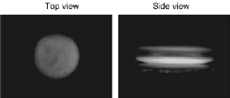
However, misuse of top view image will lead to inaccurate results concerning
the completion time of mixing operations. For example, a fluorescein droplet and
a non-fluorescein droplet are merged together for 10 s, and fluorescent microscope
capture images of the mixed droplet from top and side views, as shown in Fig.
5.10
[
31
]. From the top view, it is observed that the fluorescent reagent has been
homogeneously distributed in the merged droplet, i.e., the mixing procedure may
appear to be complete. However, from the side view, the merged droplet has multiple
layers which indicates incomplete mixing operation. Hence it is essential to monitor
cyberphysical microfluidic biochips with different perspectives. Two cameras can
be fixed onto the hardware platform so that images of the droplets can be acquired
from two different perspectives.
When multiple droplets are manipulated concurrently on the biochip, the visibil-
ity of the droplets for the side-view assessment should be considered. An example
is shown in Fig.
5.11
a. We assume that droplets 1
4 are being mixed concurrently
in four mixers on the biochip, and their directions of movement are indicated by
the arrows. We assume that the mixing operations for these four droplets start at the
same time with the droplets moving in the same direction.
AsshowninFig.
5.11
a, the initial positions of droplets 1 and 3 are in the same
row of the electrode array, and the initial positions of droplets 2 and 4 are in the same
row of the electrode array. When the movements of droplets 1 and 3 are projected on
the y-axis, they will always overlap with each other during the mixing procedure.
Therefore, if the camera is fixed at Position 1 shown in Fig.
5.11
a and we acquire
side-view images of these four droplets, droplet 1 will always be hidden behind
droplet 3, i.e., droplet 1 is “invisible” to the monitoring system. Likewise, droplet 2
will always be hidden behind droplet 4, and it is also invisible to the monitoring
system. Similarly, if the camera is fixed at Position 2 shown in Fig.
5.11
aand
monitors these four droplets from the side, droplets 2 and 4 will always be invisible
to the monitoring system.
The situation that “some of the droplets are invisible for the camera or fluorescent
microscope” must be avoided in order to monitor all of the mixing procedures on
the biochip simultaneously.
Fig. 5.10
To p
and
side views
captured after merging a
fluorescein droplet with a
non-fluorescein droplet for
10 s [
31
]