Graphics Reference
In-Depth Information
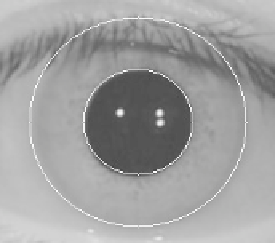
Fig. 8.
The image of outer edge of iris
4.2
Linear Segment
The sub-image also has some noises (i.e. eyelashes) which hinder the extraction of the
limbic edge. So a quarter of the up and down part has been got rid of the iris image,
only to search the middle section and look for external edge.
Here are the steps to follow:
(1)
Segment the sub-image, and the size of the rest image is 240 * 220.
(2)
Get each line elements first derivative of the rest of the image respectively.
(3)
Divide the elements of each row into three parts: the pupil and the left part and
right part of the pupil. Scanning the left part and the right part of the pupil. Be-
cause the pixel values of iris are smaller than the part of sclera, the max value is
on the left and the min value is on the right. Recording the coordinates of the max
and min values. Then some points of the outer edge are found.
(4)
Use the method of fitting to get the outer edge of the iris.
The result of the linear segment process is shown in Fig.9
Fig. 9.
Outer edge of iris by used the method of linear segment
5
Contrast Experiment
The iris database of CASIA1.0 from Chinese Academy of Sciences Institute of Auto-
mation has been used to verify the accuracy and real-time of the two methods. Results
of the different methods of the Hough, calculus, circle integral and linear
segment are
shown in Fig. 10.

Search WWH ::

Custom Search