Biomedical Engineering Reference
In-Depth Information
fluid. The latter challenge is related to the standard means of forced
cantilever oscillation, which is piezoactuation of the cantilever base; as a
result, the entire cantilever length vibrates vertically to achieve the
prescribed free-end amplitude and associated contact force. Such
oscillation close to the sample surface can generate waves within the
imaging fluid that distort the image and perturb the intended contact
force. To minimize these deleterious effects for hydrated biomaterials,
two unique hardware modifications have been developed. The first
approach employs magnetic coating of the backside of AFM cantilevers,
and actuation of the cantilever via an oscillating electromagnetic field
localized at the free-end of the cantilever. This is called magnetic AC
or MAC mode.
15
The second approach oscillates an uncoated cantilever
by driving AC current directly through the cantilever legs in the
presence of a small magnetic field; the interacting electromagnetic fields
oscillate the cantilever free-end.
16
Both approaches significantly reduce
mechanical coupling of the imaging fluid with cantilever oscillation, and
enable increased resolution in height, amplitude error, and phase lag
images from which mechanical properties can be inferred qualitatively
17
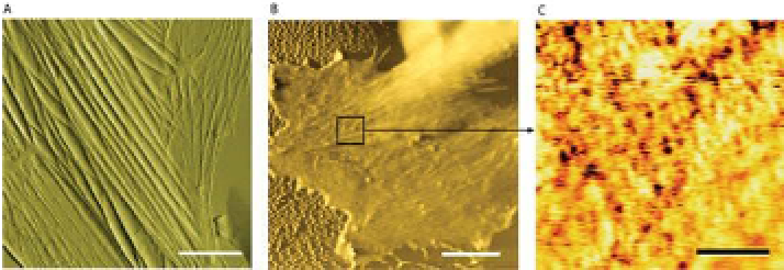
A B C
Figure 3-5. (A) Spatial resolution of living vascular endothelial cell topography via
contact mode imaging with AFM cantilevers; scalebar = 5
m. (B) Magnetic oscillation
of these cantilevers in MAC mode retains spatial resolution of cell surface, reduced here
due to fixation and stiffening of the cell itself; scalebar = 5
m. (C) Such probe
oscillation also enables spatially resolved maps of highly adhesive regions on the
cell surface, which can be demonstrated to be locations of molecular receptors; scalebar =
500 nm. (B, C).
17

Search WWH ::

Custom Search