Biomedical Engineering Reference
In-Depth Information
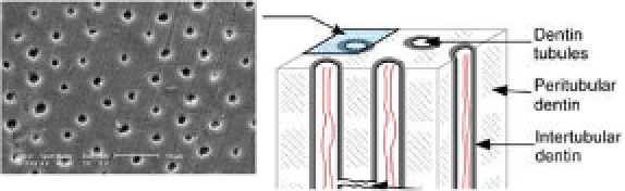
The dentin structurally consists of tubular, peritubular and
and due to the insertion of the odontoblasts process into the mineralized
From the outer surface of the dentin to the area nearest the pulp, these
tubules follow an S-shaped path. The diameter and density of the tubules
are greatest near the pulp.
26
Tapering from the inner to the outermost
surface, they have a diameter of 2.5 m near the pulp, 1.2 m in the
middle of the dentin, and 900 nm at the dentino-enamel junction. Their
density is 59,000 to 76,000 per square millimeter near the pulp, whereas
the density is only half as much near the enamel. Dentinal tubules give
high permeability to the dentin. In addition to an odontoblast process, the
tubule contains dentinal fluid, a complex mixture of proteins, such as
albumin, transferrin, tenascin and proteoglycans.
26
In addition, there are
branching canalicular systems that connect to each other to form a
complex network. Dentin tubules are surrounded by highly calcified
peritubular dentin, which is more radio-opaque and electron-dense
than intertubular dentin. The less calcified intertubular structure, which
contains more organic material than peritubular dentin, comprises the
remaining dentin body and lies between regions of peritubular dentin.
Figure 8-2. Schematic illustration of dentin tubule structure; and SEM images from
different directions, modified from Angker
et al
.
25

Search WWH ::

Custom Search