Biomedical Engineering Reference
In-Depth Information
2076.92 3095.08 4113.24 5131.40 6149.56
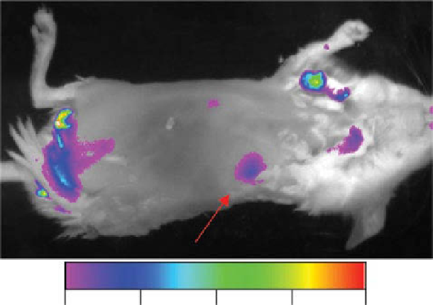
figure 3.5
Nir imaging. side view of mouse injected with Alexa-labeled micelles, at 2 h
after the injection. Arrow points the tumor. (With permission from ref. [153]. © dovepress.)
(
See insert for color representation of the figure.)
.)
of three- to four-fold in the blood pool (aorta and heart) was visually observed in rats
and rabbits for at least a period of 2 h following the injection. The quantification of
some of these images with the help of image-processing software showed no sign of
a decrease in the blood opacification during at least a 3 h period following the injec-
tion. A similar pattern was also observed in rats. micelles with such properties would
certainly offer the potential for clinical use in the diagnostic CT imaging of the blood
pool. The clinical utility of selective blood pool contrast agents may be variable:
minimally invasive angiography, image guidance of minimally invasive procedures,
oncologic imaging of angiogenesis, ascertaining organ blood volume tomographi-
cally, and identifying hemorrhage are applications that could benefit from a long-
lived intravascular agent.
diacyllipid micelles coloaded with the photosensitizer drug and iron oxide
nanoparticles have been suggested for simultaneous photodynamic therapy and mri
of tumors [146]. similar drug-/iron oxide-coloaded micelles as the possible agents
for combined chemotherapy and imaging of cancer were also suggested in [147].
general platform of target-specific micelles for molecular mri is discussed in [148].
in particular, CB2 receptor- and NgAL-targeted paramagnetic micelles were shown
to nicely image atherosclerotic plaques [149].
of special interest is the use of pEg-lipid micelles for encapsulation of quantum
dots (Qd) [150]. Qd encapsulated in lipid-based micelles have been successfully
used for visualization of tumors and metastases
in vivo
in mice with tumors
[151-153], and Qd-loaded micelles specifically targeted to tumors with antitumor
antibodies provided the best and fastest optical near-infrared images (see fig. 3.5).
Search WWH ::

Custom Search