Biomedical Engineering Reference
In-Depth Information
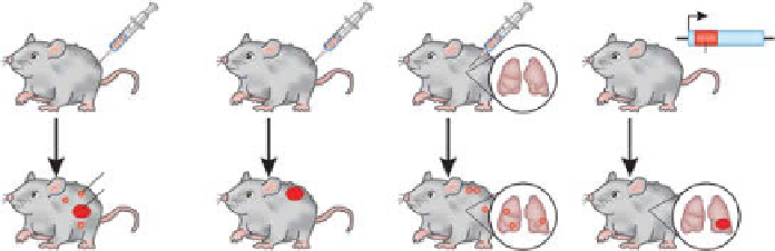
Subcutaneous
xenograft
-Transplanted
-Immunodecient
Orthotopic
xenograft
-Transplanted
-Immunodecient
Syngeneic
-Transplanted
-Immunocompetent
GEMM
-Spontaneous
-Immunocompetent
KRAS
G12D
Metastasis
Primary tumor
Metastatic
Nonmetastatic
Metastatic
Nonmetastatic
figure 16.4
Cartoon of common mouse used in biomedical research. Source: Katie Vicari.
(Reprinted with permission from Ref. [14]. © Macmillan Publishers ltd.)
16.4.2.3 Xenografts
Early research in cancer required models in which cancer
development could be easily assessed, such as the subcutaneous xenografts. The
most commonly used animal models of cancer are the human cancer xenografts in
immunocompromised mice. For optical imaging, the nude mouse is particularly
well suited as these strains are relatively hairless, which benefits optical imaging.
They are also sufficiently immunocompromised to allow growth of many human
cancer cell lines. Severe combined immunodeficiency (SCID) mice are more immu-
nocompromised and allow growth of more cell lines and human tissues but have
hair. In these models, tumors grow after implantation of human cancer cells from
immortalized cell cultures or transplanted tissues. Implantations can be done ectop-
ically such as underneath the skin (subcutaneous) or in the tissue of origin (ortho-
topic). Subcutaneous xenografts with human cancer cell lines are relatively simple
to perform and enable large cohorts for experiments. Subcutaneous xenografts have
been called “walking petri dishes” because of the relatively homogenous growth and
lack of stromal tissues and appropriate architecture compared with natural human
disease. Also, of particular importance to molecular imaging, vasculature tends to be
more chaotic and leakier than in human disease, leading to EPR effects and higher
nonspecific accumulation.
Bilateral tumor xenografts can provide additional sample numbers for tumors if the
same cell lines are implanted. Different cell lines can be implanted for direct comparison
in the same animal. For example, the M21 melanoma cell line expresses high levels of
alpha-v-beta-3 (avb3) integrin receptors, while the M21l expresses very low levels
of avb3. These cell lines have been implanted in opposite flanks of nude mice for
molecular imaging studies of molecular probes targeting avb3. The difficulty with this
approach is that the tumors grow at different rates and implantation time must be
varied accordingly. It is also likely that other aspects of tumor biology may be different
in these tumors as avb3 is associated with angiogenesis. Vascularization and vascular
permeability may be altered. These and other differences must be accounted for in
imaging studies.
Search WWH ::

Custom Search