Biomedical Engineering Reference
In-Depth Information
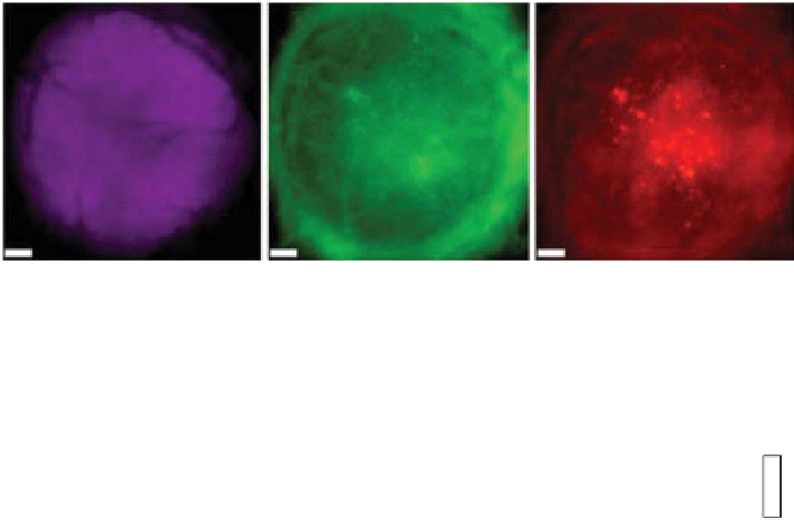
(a)
Tumor
CPMV
PVX
(b)
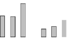
60
53
50
40
33
32
27
30
16
20
13
7
5
4
10
0
PBS
PVX
CPMV
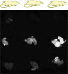
figure 14.4
images of tumor homing of pVX versus CpMV in human tumor xenograft
models. (a) intravital imaging of the CAM model. Chick embryos with human epithelial carci-
noma hep3 tumors (magenta) were coinjected with equal numbers of particles of Alexa Fluor
555-labeled pVX (pVX-A555) and Alexa Fluor 647-labeled CpMV (CpMV-A647). imaging
was performed 4 hours after injection. Scale bar is 190 µm. (b) Fluorescent images of hT-29
tumors excised from mouse tumor xenograft model. pVX-A647 and CpMV-A647 were
injected by tail vein, and tumors were excised and imaged 24 h after injection (left). Fluorescence
intensities of the tumors were then quantified (right). (reprinted with permission from ref.
[40]. © American Chemical Society.)
14.5
moleculAr TArgeTed fluorescence imAging
Several VNp formulations have been designed as active cancer-targeting optical
imaging agents. A number of advances have been made to link targeting moieties to
deliver VNps containing imaging/contrast agents to specific disease sites while avoid-
ing healthy tissues. Targeted imaging increases the signal-to-background ratio to allow
more accurate diagnosis. VNps provide a valuable platform for the development of
imaging tools due to their high versatility for chemical bioconjugation. Combinatorial
library approaches, including phage display and one-bead-one-compound (oBoC)
libraries, have pioneered the discovery of tumor-specific ligands and vascular homing
peptides to facilitate and advance the development of targeted VNps [82-87].
The following sections highlight just a few of many examples of the utility of
VNps displaying specific ligands for targeted imaging. one method for cell targeting


Search WWH ::

Custom Search