Biomedical Engineering Reference
In-Depth Information
(a)
(b)
(c)
Cell cycle
Rayleigh images
Raman spectra
3 h
4 h
5 h
6 h
7 h
8 h
9 h
10 h
11 h
12 h
G1
3 h
10 h
S
16 h
13 h
14 h
15 h
16 h
17h
18 h
19 h
20 h
21 h
22 h
23 h
24 h
G2
21 h
M
23 h
G1
400
600
800 1000
Wavenumber (cm
-1
)
1200
1400
1600
24 h
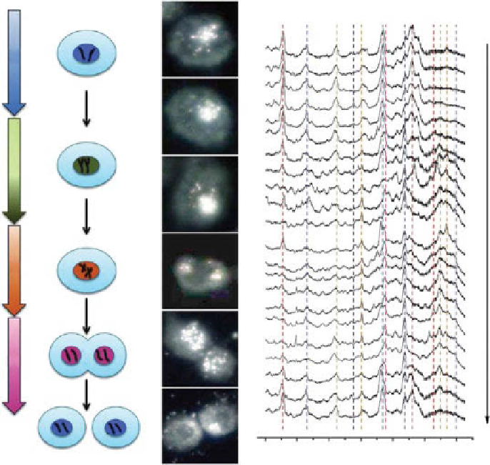
figure 11.4
Real-time dark-field images and spectra of the targeted nucleus during the
complete cycle of a single HSc-3 cell: (a) cell cycle, (b) plasmonically enhanced Rayleigh
images, (c) corresponding SERS spectra showing that different phases of the cell cycle could
be identified based on the Raman peaks attributed to Dna and protein. (Reprinted with per-
mission from Ref. [31]. © american chemical Society.)
SERS-based bioimaging. ideally, SERS probes intended for
in vivo
bioimaging
should (i) provide large, uniform, and stable SERS signal with niR excitation (650-
900 nm), (ii) be biocompatible and stable in complex physiological fluids and
exhibit long blood circulation time, and (iii) be amenable for biofunctionalization
for targeted delivery. The key considerations in the design of such efficient SERS
probes include the following.
11.4.2.1 Composition of Plasmonic nanostructures
along with other factors
such as size and shape of nanostructures, composition (of both bulk and surface) of
plasmonic nanostructures plays a determining role in the biocompatibility of SERS
probes for
in vivo
applications. Silver nanoparticles exhibit stronger plasmon fields
than gold nanoparticles of same shape and size based on the fact that the plasmon
Search WWH ::

Custom Search