Biomedical Engineering Reference
In-Depth Information
(a)
(b)
Coll
M
Ex
D
(c)
Fluorescence
Raman
Hand-held
“SpectroPen”
Optical
ber
Spectrometer/laser
5 m
10 cm
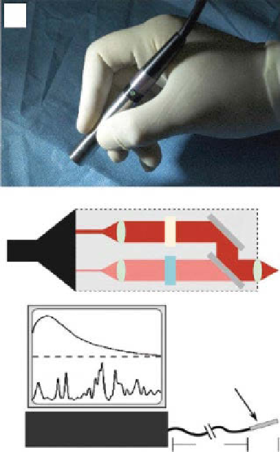
figure 11.2
(a) Photograph showing the SpectroPen held by an operator in a surgical setting.
(b) optical beam paths of the SpectroPen. a 785 nm laser diode (200 mW output) serves as an
excitation source. The illustration also shows excitation fiber (Ex), band-pass filter (BP), dichroic
filter (D), collection fiber (coll), long-pass filter (LP), and reflective mirror (M). (c) Schematic
illustration of the hybrid spectrometer designed for wavelength-resolved fluorescence and Raman
scattering. (Reprinted with permission from Ref. [21]. © american chemical Society.)
called SpectroPen that enables wavelength-resolved fluorescence and Raman
measurements for
in vivo
and intraoperative SERS-based bioimaging applications, as
showed in figure 11.2 [21]. This SpectroPen device effectively removes silica Raman
peaks from the fiber optics and reflected excitation light, allowing resolvable
fluorescence and Raman signals when contrast agents were buried 5-10 mm deep in
fresh animal tissues. after surgery, the SpectroPen device provides further evaluation
of both positive and negative tumor margins around the surgical cavity, raising new
possibilities for real-time tumor detection and image-guided surgery.
11.3.3.2 Seeing through Diffuse Human Tissue
a different line of efforts is
focused on enhancing the depth of imaging. it is known that commonly employed
backscattering geometry suffers from limited depth of penetration (only several
Search WWH ::

Custom Search