Biomedical Engineering Reference
In-Depth Information
contrast agents [140, 141]. due to the frequencies (on the order of MHz) of clinical
US imaging systems, the sizes of these bubbles are on the order of micrometers.
Since typical microbubbles do not intrinsically absorb light because of its optical
transparency, additional PA contrast agents were added into microbubbles.
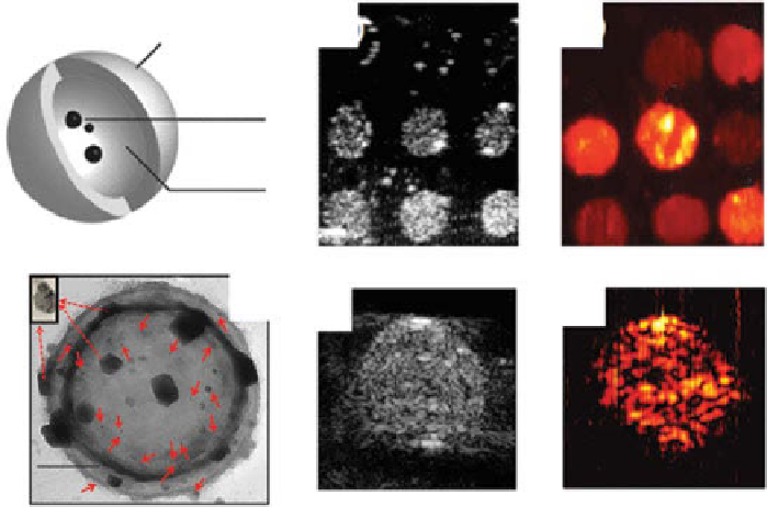
Figure 10.10a shows the schematic of encapsulated-ink poly(lactic-co-glycolic
acid) (PLgA) microbubbles and nanobubbles [138, 142]. The average sizes of
microbubbles and nanobubbles were approximately 1 and 0.3 µm, respectively.
Figure 10.10b and c are US and PA images obtained from a tissue-simulating
phantom, respectively. objects 1 through 4 were composed of encapsulated-ink
microbubbles at concentrations of 2.5, 5.0, 10, and 15 mg/ml, respectively, while
objects 5 through 8 were made of encapsulated-ink nanobubbles at concentrations
of 2.5, 5.0, 10, and 15 mg/ml, respectively. All eight objects are clearly shown in
both the US and PA images. Linear increases of the US and PA signals were observed
as a function of concentration. As the second example, Figure 10.10d shows the
TEM image of encapsulated gold nanorod human serum albumin (HSA)-shelled
microbubbles (Aunr-HSA) [139]. The US and PA images acquired from a sample
containing Aunr-HSA are shown in Figure 10.10e and f, respectively. As another
example, liquid perfluorocarbon (PFC) nanodroplets with encapsulated plasmonic
2
2
1
1
(c)
(b)
PLGA shell
(a)
5
3
4
3
4
5
India ink
6
7
8
8
Air
6
7
Assembly of AuNR-HSA
(d)
(e)
(f)
500 nm
figure 10.10
dual-modality photoacoustic (PA) and ultrasound (US) contrast agents.
(a) Schematic of PLgA microbubbles (MBs) and nanobubbles (nBs) encapsulating black
india ink. (b) US and (c) PA images of a phantom containing tumor simulators made of encap-
sulated-ink MBs and nBs with various concentrations. 1 through 4: MBs at concentrations of
2.5, 5.0, 10, and 15 mg/ml, respectively. 5 through 8: nBs at concentrations of 2.5, 5.0, 10, and
15 mg/ml, respectively. (d) TEM images of albumin-shelled MBs with encapsulated gold
nanorods (AuMBs). (e) US and (f) PA images of a phantom containing AuMBs. (reprinted
with permission from refs. [138, 139]. © SPIE.)
Search WWH ::

Custom Search