Biomedical Engineering Reference
In-Depth Information
10.4
intrinsic contrasts for Pat
PAT has been successfully provided anatomical and functional information of
biological tissues using intrinsic contrasts. Major endogenous absorbents include
two types of hemoglobin (i.e., oxyhemoglobin and deoxyhemoglobin), melanin,
lipid, and water [80]. In addition, by using different optical wavelengths, PAT can
spectrally distinguish each chromophore. For example, two types of hemoglobin and
melanin dominantly absorb visible light, while water and lipid absorb near-infrared
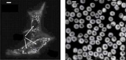
(nIr) light. Figure 10.4 shows the agent-free morphological PA images. A single
melanoma (Fig. 10.4a) and red blood cell (Fig. 10.4b) are clearly imaged by using a
subwavelength optical-resolution PAM system
in vitro
, which confirms the capability
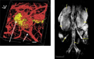
of agent-free PA imaging [81]. As shown in Figure 10.4c, PAT clearly visualizes the
morphologies of a B16 melanoma skin cancer and its surrounding vasculatures in
small animals
in vivo
based on these intrinsic contrasts [76]. Further, PAT can
provide the whole-body small animal images acquired with endogenous contrasts
and an array-based PACT system. Figure 10.4d shows volumetric PA rendering of
(a)
(b)
5 µm
10 µm
CN
(c)
(d)
Spleen
Partial lobe
of liver
Right
kidney
Left
kidney
Ovarian
vessel
Melanoma
BV
3 mm
Ovarian
vessel
Interior
vena cava
5 mm
figure 10.4
PA images of intrinsic chromophores.
In vitro
PA images of (a) B16 melanoma
cells and (b) red blood cells. Cn, cell nuclei (reprinted with permission from ref. [81].
© optical Society of America.), (c) Volumetric anatomical PA image of B16 melanoma
(778 nm) and its feeding blood vessels (570 nm) (reprinted with permission from ref. [76].
© American Chemical Society.). BV, blood vessels, (d)
In vivo
noninvasive volumetric PA
image of the whole nude mouse body obtained at 755 nm. (Permission from ref. [71]. © SPIE.)
Search WWH ::

Custom Search