Biomedical Engineering Reference
In-Depth Information
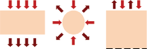
(a)
(b)
(c)
FIgure 9.9
Different imaging probe configurations, (a) transmission mode, (b) transmission
and reflection mode, and (c) reflection mode.
example, temperature and pH, or molecular interactions [40, 41]. on the other hand,
its value does not depend on the concentration of the fluorophores or the intensity of
the excitation light (before saturation) [28]. The sensitivity of fluorescence lifetime
to biochemical environment is based on the structure of the dye. To evaluate specific
properties of the probe environment in tissue, it is important to choose a proper dye
with lifetime sensitivity that is dominated by only one specific factor.
In terms of imaging probe geometry, light can transmit through the tissue in reflec-
tion mode, in transmission mode, or in both reflection and transmission mode
(Fig. 9.9). In transmission mode (transillumination), the source and detector are placed
on opposite sides of the tissue; and the detector captures the transmitted photons
through the tissue. In reflection mode (epi-illumination), the source and detectors are
placed on the same side of the tissue, and the reflected fluorescence signal from
the tissue is captured. In many medical applications, using transillumination is not
possible, and the source and detectors have to be placed on the same side of the object.
It is also possible to position the source and detectors (e.g., in a ring geometry) so that
the fluorescence signal can be captured in both reflection and transmission mode.
To detect the fluorescence signals coming from different depths in epi-illumination
mode, multiple sources and detectors are required to be positioned at different distances.
This is due to the banana-shaped pattern of light propagation in high scattering media
(Fig. 9.8). As the distance between source and detector increases, the center of the path
covers a deeper area inside the tissue. owing to the diffusion of excitation and emission
light in tissue, epi-illumination can image fluorescence activities at depths ranging
from millimeters to a few centimeters, based on the distance between sources and
detectors and the excitation and emission wavelengths of the fluorophore.
Data can be collected either by scanning of the roI by the source-detector pairs
or by the area detection, using a camera. A mechanical scanner or a mirror with
galvanometer is usually used to scan the roI.
The most common fluorescence imaging scheme is planar imaging. This method
projects all the fluorescence light on the tissue surface without considering the depth
of fluorescence target. It is fast and easy to implement and works very well on
superficial fluorescence targets. However, in deep tissue imaging, even though the
fluorescence intensity is a linear function of fluorophore concentration, it depends
nonlinearly on optical properties of tissue and the fluorophore depth inside the media.
Search WWH ::

Custom Search