Biomedical Engineering Reference
In-Depth Information
1.00
O
2
Hb (mm
-1
mM
-1
)
HHb (mm
-1
mM
-1
)
H
2
O (mm
-1
×20)
Bulk lipid (mm
-1
× 50)
0.75
0.50
0.25
0.00
650
700
750
800
850
900
950
1000
Wavelength (nm)
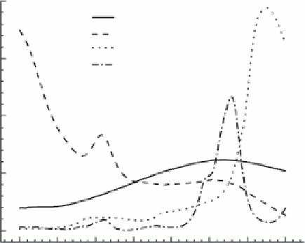
FIgure 9.4
Absorption spectra of main chromophores in biological tissue in the NIr
window. (reprinted with permission from ref. [37]. © IoS Press.)
(a)
(b)
FIgure 9.5
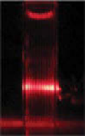
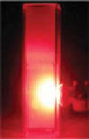
light propagation in (a) nonscattering and (b) high scattering media.
9.3.2
scattering
In nonscattering media, light propagates along straight lines. Therefore, any occlu-
sion in the media can be imaged or reconstructed with a simple projection or back
projection algorithm similar to X-ray or computed tomography (CT). However, due
to the highly scattering nature of biological tissues, photons change their propagation
directions randomly throughout the media, and finding the occlusions inside the
media is not straightforward as in the case of nonscattering media. Figure 9.5 shows
the light propagation path in (i) nonscattering and (ii) high scattering media. random
propagation of light in high scattering media significantly decreases the resolution
in the deep tissue images.
The light source can illuminate the media in different modes, that is, a continuous-
wave (CW), short pulse, or frequency-modulated signal. The effect of scattering
media on a CW is the same as absorption and attenuates the received photons to the
detector. The photons propagate in all directions inside the media, and not all of them
can reach the detector.
Search WWH ::

Custom Search