Biomedical Engineering Reference
In-Depth Information
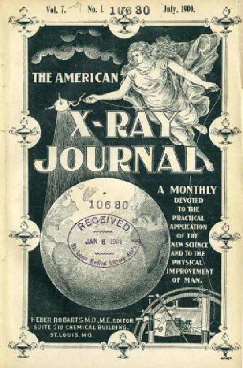
figure 1.3
The
American X-Ray Journal
established in May 1897 was one of the first
imaging journals. launched by Dr. H. robarts, a prominent radiologist from St. louis, his
biography is described in ref. [2]. The journal existed until 1905. (courtesy of Becker library,
Washington University School of Medicine.)
processes of digestion, the movements of the food in the stomach and small intestine
were observed by means of the shadows cast on a fluorescent screen” [3]. a few
years later, a less toxic barium sulfate mixed with foodstuffs became the first broadly
used contrast agent in X-ray imaging of the digestive tract [4]. This water-insoluble
salt (to prevent barium toxicity) was swallowed with food prior to the imaging
procedure to outline the esophagus, stomach, and small intestines. The contrast could
also be inserted via enemas to visualize the colon. This practice allowed the visuali-
zation of tumors, strictures, blockages, and ulcers and has been so simple and suc-
cessful that it is still in use today.
The next advancement in the development of contrast agents came from argentina,
where in 1919 the radiologist Dr. c. Heuser intravenously injected a water-soluble
Search WWH ::

Custom Search