Biomedical Engineering Reference
In-Depth Information
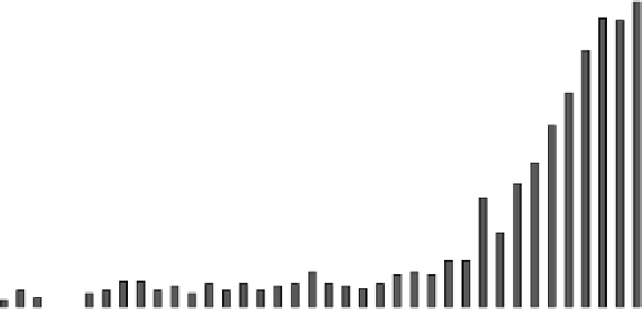
3.50
3.00
2.50
2.00
1.50
1.00
0.50
0.00
figure 1.1
growth of the nanoparticle research in biomedical imaging. Solid arrows
show the appearance of imaging techniques, and dotted arrows show the emergence of
nanoparticles. a number of citations are given from PubMed database.
into a mature field known today as molecular imaging. figure 1.1 reflects a remarkable
tenfold increase in nanoparticle-related medical imaging research from a relatively
modest approximately 0.25-0.3% in the twentieth century to the current 3%. This
growth resulted in more than 1500 nanoparticle imaging-related publications in
2012 alone.
from the onset of radiology and the first contrast agents to the end of the
twentieth century, imaging techniques such as X-ray, PeT, SPecT, ultrasound,
MrI, optical, and photoacoustics have emerged. The first imaging nanoparticles
appeared only in the middle of the twentieth century. The progress and the appli-
cation of imaging nanoparticles followed the advent of new imaging modalities
and diverged into two equally important directions. In one direction, de novo
nanoparticle designs were developed for specific imaging modalities. Some exam-
ples include magnetic particles for MrI, quantum dots (QDs) for optical, and
nanobubbles for ultrasound. The other direction adopted previously established
designs of nanoparticles (for instance, for drug delivery) and modified them for
imaging applications. Some examples include liposomes, virions, cross-linked
nanoparticles, and surface modification to increase the nanoparticles' imaging
specificity. regardless of direction, many nanoparticles applications often began
as unexpected discoveries. Many steps to refine their design were necessary to turn
them from a mere curiosity to a clinically acceptable tool. Today, the continued
improvement in nanoparticle synthesis, conjugation technique, and novel bio-
markers made the nanoparticle approach a unique and well-differentiated scientific
direction that blends seamlessly with clinical imaging. The historical trend illus-
trated in figure 1.2 highlights the most important milestones toward this direction
and is discussed in this chapter.


Search WWH ::

Custom Search