Biomedical Engineering Reference
In-Depth Information
contrast agents such as hemoglobin and melanin [18]. These and some other endogenous
molecules have much stronger light absorption properties in the NIR region than the
surrounding tissue; hence, the intensity of an OA signal in biological tissue is propor-
tional to optical energy absorption of the contrast agent [31]. endogenous OA con-
trast agents are usually not targeted when used, but they can be incorporated into the
matrix of nanoparticles with specified binding of molecules of interest. examples
include hemoglobin and melanin, the two most important naturally occurring con-
trast agents for enhanced OA imaging.
5.2.1
hemoglobin
As an endogenous contrast agent, hemoglobin has been most widely explored for OA
imaging and tomography. OA imaging with hemoglobin was shown to greatly facil-
itate blood dynamics related to brain research [32-34] and help with visualizing
brain structure and lesions [32], delineating tumor vasculature [35], monitoring
hemodynamics, measuring microvascular blood flow [36], and imaging whole body
of animals
in vivo
and
ex vivo
[37, 38].
OA noninvasive visualization of the brain's vascular system could be of great
importance for studying the function of the brain, diagnosing possible disorders, and
providing clinically translatable insights into the progression of various human neu-
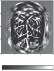
rological diseases. The structures of the surface of a rat brain with hemoglobin as
contrast agent were mapped with OA imaging (fig. 5.1) by Wang's group from the
university of michigan, school of medicine. The report [32] describes the functional
cerebral hemodynamic changes in cortical blood vessels around the whisker-barrel
cortex evaluated in response to their stimulation, as well as hyper- and hypoxia-
induced cerebral hemodynamic changes. Other reports from the same research group
0
0.5
1.0
(cm)
1.5
2.0
0
0.5
1.0
(cm)
1.5
2.0
0
0.5
1.0
(cm)
1.5
2.0
Min Differential absorption (Δ
A
) Max
Min Differential absorption (Δ
A
) Max
Min Optical absorption (
A
) Max
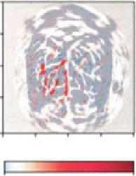
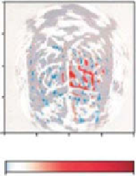
figure 5.1
Nontargeted functional OA imaging of cerebral hemodynamic changes in
response to whisker stimulation. Left: OA image of the vascular pattern in the superficial layer
of the rat cortex acquired with the skin and the skull intact. middle: Noninvasive functional
OAT images corresponding to left-side whisker stimulation. Right: Noninvasive functional
PAT images corresponding to right-side whisker stimulation. (Adapted with permission from
Ref. [32]. © macmillan Publishers Ltd.)
Search WWH ::

Custom Search