Biomedical Engineering Reference
In-Depth Information
(a)
(b)
200 nm
5 min
1h
(c)
(d)
Intensity
Lifetime
0.72
0.65
5 min
1h
5 min
1h
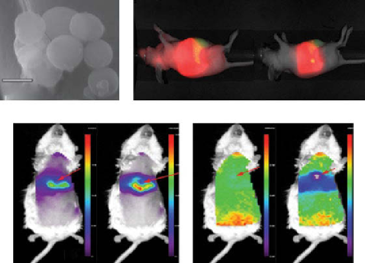
Figure 4.20
(a) SEm images of NCs with ICg after treatment with 4% w/v BSA. (b)
Fluorescence intensity after administration of ICg-NCs. Red (ICg) channel emission (800 nm)
and green channel emission (700 nm) correspond to endogenous fluorescence (from the diet).
Red fluorescence is primarily localized in the liver. (c) Intravenous (IV) injection of ICg-NC
shows an accumulation of the fluorescent dye in the liver. Left, fluorescence intensity; right,
fluorescence lifetime, ex/em = 775/820 nm. (
See insert for color representation of the figure.)
polymersome poration compared with the extracellular environment. Temperature
was also found to have an effect on the rate of polymersome poration.
A near-infrared fluorophore (NIRF)-labeled peptosome was prepared by adding
ICg-conjugated polypeptide. Diameter of the peptosome prepared from gA-PSar-
PmLgs was in the range from 100 to 200 nm, which is the appropriate size for the
passive targeting into the solid tumor by the EPR effect.
In vivo
retention in the blood
of ICg-labeled peptosome was assayed by using rats as experimental animals, and
the retention time was proven to be as long as the PEgylated liposome. ICg-labeled
peptosome was also administered to the tumor-bearing mouse and successfully
detected the tumor tissue due to the EPR effect [88].
The anticancer drug DOX and hydrophilic superparamagnetic iron oxide nanopar-
ticles (SPIONs) were encapsulated inside the inner aqueous core of the vesicles for
cancer therapy and mRI, respectively. It was shown that consequently effective sup-
pression of cancer cell growth was detected [89].
Novel functional hollow chitosan-poly(acrylic acid)-gold (CS-PAA-Au) hybrid
nanospheres, prepared in an aqueous solution via a one-pot route, can not only act as
Search WWH ::

Custom Search