Chemistry Reference
In-Depth Information
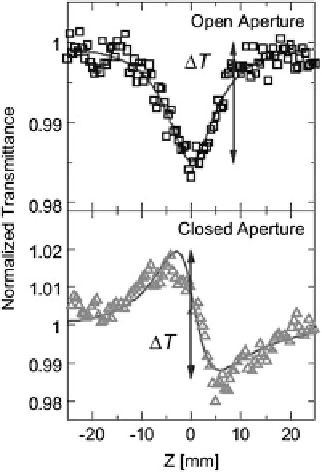
Fig. 7.11 Typical Z-scan
profiles of the [Ni(
L
)
2
Br]Br
2
/
PMMA film in the open and
closed aperture condition.
The
solid lines
show the
theoretical fits. Adapted
from [
5
]
image in Fig.
7.10a
. The photograph of the film on a CaF
2
substrate is shown in
Fig.
7.10b
.
Figure
7.10c
shows the absorption (
) spectrum of the [Ni(
L
)
2
Br]Br
2
film,
together with the spectrum of a single crystal of [Ni(chxn)
2
Br]Br
2
. The latter is
obtained as the sum (
a
a
⊥
, which
were calculated from the polarized reflectivity spectra for the electric field of light
parallel and perpendicular to
b
, respectively, through the KK transformation. The
spectral shape of
a
//
+2
a
⊥
) of the polarized absorption spectra
a
//
and
in the [Ni(
L
)
2
Br]Br
2
film is in good agreement with that in the
[Ni(chxn)
2
Br]Br
2
single crystal. No significant enhancement of the background due
to light scattering is observed even in the higher energy region (~4 eV), suggesting
the high quality of the film as an optical media.
Typical Z-scan profiles of the [Ni(
L
)
2
Br]Br
2
film are presented in Fig.
7.11
. The
measurements were carried out with the light pulse of the optical communication
wavelength
a
l ¼
m
ho ¼
:
80 eV ). The upper panel
shows the nonlinear increase in the absorption (decrease of the transmittance)
around the focal plane (
z ¼
1.55
m (the photon energy
0
0) in the open aperture condition. The one-photon
absorption is negligible at 0.8 eV so that the observed nonlinear signal can be
attributed to two-photon absorption (TPA). The profile in the partially closed
aperture condition includes the TPA component as well as the component for the
optical Kerr effect described by Re
w
ð
3
Þ
ðo; o; o; oÞ
. By dividing the profile in
the partially closed aperture condition by that in the open aperture condition, the
profile for the optical Kerr effect alone was obtained, which exhibits a plus-minus
structure characteristic of self-defocusing, as shown in the lower panel of Fig.
7.11
.
The two profiles in Fig.
7.11
were well reproduced by the theoretical profiles
predicted for the third-order nonlinear optical response, as shown by thin solid
Search WWH ::

Custom Search