Biomedical Engineering Reference
In-Depth Information
(b)
(a)
3940
3650
3360
3070 2780
Wavenumber (cm
-1
)
2490
2200
1910
1620
1330
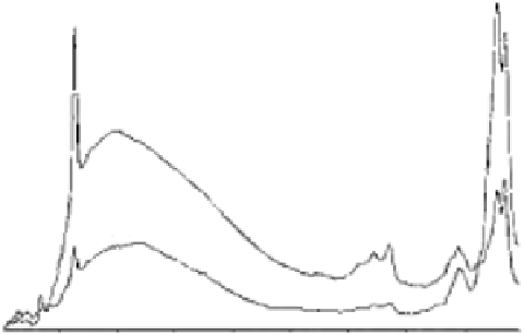
Figure 7.15
FTIR spectra of carbonated (a) and hydroxyl (b) apatites, carbonate and hydroxyl bands. (With
permission.)
seldom seen in the infrared spectrum [20,21]. Usually, ν
1
and ν
4
have strong
vibrational bands in the Raman spectra of bone and carbonated apatite [22,24],
and ν
2
and ν
3
vibrational modes are observed in the infrared spectra. Carbonate
ions occupy two different sites in carbonated apatite: peaks in the region of
1650 to 1300 cm
−1
are due to the ν
3
vibrational mode carbonate ion and the peak
at 873 cm
−1
is due to the ν
2
vibrational mode [29]. These carbonate bands in the
region of 1650 to 1300 cm
−1
are assigned to surface carbonate ions, rather than
to carbonate ions in the lattice of phosphate ions. The ν
3
has a peak split in two
peaks centred at 1649 and 1470 cm
−1
, respectively, with the distribution of the
carbonate ν
3
sites depending on the maturation and formation of apatite crys-
tals. Occupancy of the ν
2
sites is considered to occur competitively between the
OH
−
and carbonate groups at the interface of growing crystal, whereas occu-
pancy of the ν
3
sites depends on competition between the phosphate and car-
bonate ions [20,30]. This presence of ν
2
and ν
3
vibrational modes of carbonates
in carbonated apatite may contribute to the decrease of hydroxyl ions in the
spectrum, as the hydroxyl band present in the spectrum of carbonated apatite
is weaker in intensity than that in commercial HA powders.
The spectra of synthetic hydroxyapatites also have peaks in the region of
1650 and 1300 cm
−1
. Carbonated apatite has two well-defined peaks for the ν
3
sites centred at 1649 and 1470 cm
−1
, whereas synthetic commercial hydroxyapa-
tite has three sites for ν
3
vibrational mode centred at 1648, 1454, and 1419 cm
−1
.
Results obtained by peak area calculation of the carbonate ν
3
band indicate
that carbonated apatite has more carbonate moiety than hydroxyapatite. The
peak areas of carbonated apatite and hydroxyapatite powders are 165 and 13,
respectively. The spectra of the carbonated bands of carbonated apatite and
hydroxyapatite powders are compared in Figure 7.15.

Search WWH ::

Custom Search