Biomedical Engineering Reference
In-Depth Information
3 Structure and Mechanism of Action of N-Type
Voltage-Gated Ca
2+
Channels
Neuronal NCCs are complex protein machinery with several different distinct
regulatory subunits composed of
a
1
,
a
2
-
d
,
b
, and
g
subunits. NCC is mainly
encoded by Cav2.2 gene, the pore forming
a
1B
subunit and which in turn contains
the ligand sensing residues. This subunit is essential for NCC functions, and which
in turn control the channel properties.
a
1B
subunit is primarily expressed in the
neuron of spinal cord, that is at the terminals of peptidergic primary afferent
neurons and projecting to the superficial laminae of its dorsal horn [
32
].
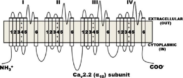
The
a
1
subunit pore with molecular mass of 190 kDa is the primary hydrophobic
subunit gene necessary for channel functioning, and it incorporates the conduction
pore, the voltage sensor and gating apparatus, with known sites of channel regula-
tion by second messengers, drugs, and toxins [
33
]. This subunit is characterized by
four homologous I-IV domains having six transmembrane helices (S1-S6) each.
The voltage sensor of the channel lies at S4 segment and each third and fourth
amino acid of this segment contains positive arginine and lysine residue [
34
].
Transmembrane segments S5 and S6 in each domain have different pore loops,
which controls its ion conductance and selectivity. In each pore loop due to the
negatively charged residues such as glutamate is exquisitely Ca
2+
selective. The
channel pore called P-region is created by four glutamate residues located centrally
in the Ca
2+
channel of
a
1
subunit gene. The movement of Ca
2+
between pore and
cell occurs, that is in the middle of the pore. Ca
2+
is then shifted into the cell after
binding of second Ca
2+
in the pore region. The auxiliary subunits
a
2
-
d
,
b
and
g
subunit genes present in these ion channels modulate the properties of the NCC
complex [
35
,
36
] (Fig.
1
).
1B
subunit of voltage-gated Ca
2+
channels. The pore-forming
Fig. 1 Schematic representation of
a
a
1B
subunit has four repeat domains (I-IV) connected by linker regions, each repeat domain has
six transmembrane segments (1-6)