Biomedical Engineering Reference
In-Depth Information
(a)
TEOS
Calcination
Si
Si
Si
(b)
(d)
(a)
PVP
(c)
CTAB
(b)
(c)
200 nm
(d)
(e)
100 nm
50 nm
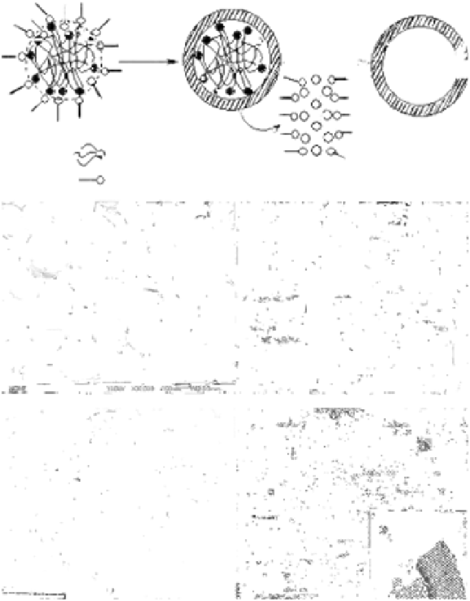
FIGURE 3.1
(a) Schematic drawing of the formation process of HMS nanoparticles. (b) SEM image of
HMS nanoparticles. (c) TEM image of the as-synthesized HMS nanoparticles. (d) TEM and (e)
HRTEM images of the calcined HMS nanoparticles. (Reprinted from Zhu Y., Shi J., Chen H., et
al.,
Micropor. Mesopor. Mater
. 84: 218-222, 2005, with permission from Elsevier Ltd.)


Search WWH ::

Custom Search