Biomedical Engineering Reference
In-Depth Information
(a)
(b)
β-TCP
β-TCP
Akermanite
NB
NB
β-TCP
NB
NB NB
NB
Akermanite
(c)
(d)
NB
NB
NB
NB
NB
β-TCP
NB
NB
Akermanite
β-TCP
β-TCP
NB
Akermanite
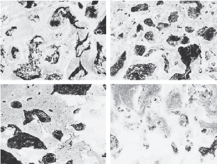
FIGURE 2.8
(
See color insert.
) High magnification images of new bone formation and material degrada-
tion of (a, c) akermanite and (b, c) β-TCP implants after (a, b) 8 and (c, d) 16 weeks (Van Gieson's
picrofuchsin staining of transverse section; NB: new bone). Red color indicates newly formed
bone. Original magnification: 100×.
Akermanite scaffolds were implanted into rabbit femur defect models and
the results indicated that both in early- and late-stage implantations, aker-
manite promoted more osteogenesis and biodegradation than did β-TCP
(Wu and Chang, forthcoming); and in late-stage implantations, the rate of
new bone formation was faster in akermanite than in β-TCP as shown in
Figure 2.8 (Huang et al. 2009). The akermanite ion extract predominantly
promoted the proliferation of human aortic endothelial cells and upregu-
lated the expression of genes encoding the receptors of proangiogenic
cytokines and the expression level of genes encoding the proangiogenic
downstream cytokines, such as nitric oxide synthase and nitric oxide syn-
thesis. Akermanite implanted in the rabbit femoral condyle model promoted
neovascularization after 8 and 16 weeks of implantation, which confirmed
its stimulation effect on angiogenesis
in vivo
(Zhai et al. 2012).
Recently, 1-mm baghdadite ceramic spheres were implanted into the supra-
condylar site of the femur defects in Wistar rats and the degree of
in vivo
osteogenesis was evaluated by hematoxylin and eosin, Safranin O staining,
tartrateresistant acid phosphatase (TRAP) staining, and immunohistochem-
istry (type I collagen: Col I; osteopontin: OPN) analyses. The results have
Search WWH ::

Custom Search