Biomedical Engineering Reference
In-Depth Information
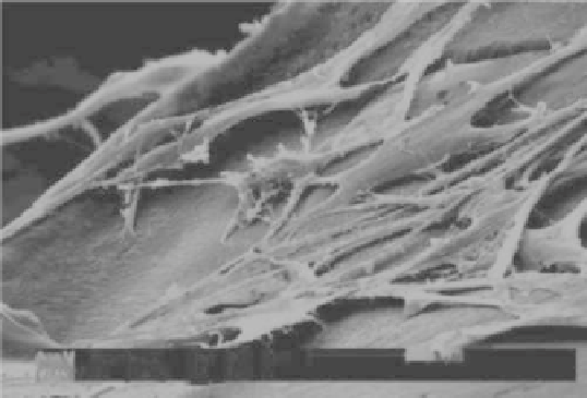
FIGURE 1.8
BMSCs morphology on MBG scaffolds after culturing for 7 days.
Our recent study revealed that the incorporation of Co
2+
ions into MBG
scaffolds significantly enhanced VEGF protein secretion, hypoxia-induc-
ible factor (HIF)-1α expression, and VEGF gene expression of BMSCs.
The incorporation of Co into MBG scaffolds is an efficient way to pre-
pare hypoxia-mimicking tissue engineering scaffolds with significantly
improved hypoxia function (Wu, Zhou, et al. 2012). The incorporation of
Sr into MBG scaffolds has significantly stimulated the proliferation, ALP
activity, and osteogenesis- and cementogenesis-related gene expression
of periodontal ligament cells. The results suggested that bioactive ions
released from MBG play an important role in enhancing cell response and
their biological functions.
1.4.3
In Vivo
Osteogenesis of MBG
To investigate the
in vivo
osteogenesis of MBG, MBG particles were
implanted into the defects of rat femur. After 8 weeks of implantation, MBG
particles induced a great amount of new bone ingrowths in the defects (see
Figure 1.9). Furthermore, MBG particles were incorporated into silk scaffolds
and investigated the
in vivo
osteogenesis. The study showed that MBG/silk
scaffolds induced a higher rate of type I collagen synthesis and new bone
formation after implanted in rat calvarial defects compared to conventional
NBG/silk scaffolds. The results confirm that MBG has significant capacity
to improve the
in vivo
bioactivity of silk scaffolds (Wu, Zhang, et al. 2011).
The preliminary results indicate that MBG has excellent
in vivo
osteogenesis
for potential bone repair application.

Search WWH ::

Custom Search