Biomedical Engineering Reference
In-Depth Information
500 µm
500 µm
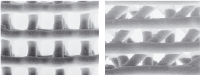
FIGURE 1.4
3D-printed MBG scaffolds with controllable pore architecture.
uniform pore structure, their mechanical strength is compromised because
of the incorporation of methycellulose, which results in some micropores. A
new facile method was recently used to prepare hierarchical and multifunc-
tional MBG scaffolds with controllable pore architecture, excellent mechan-
ical strength, and mineralization ability for bone regeneration application
by a modified 3D-printing technique using polyvinyl alcohol (PVA) as a
binder. The obtained 3D-printing MBG scaffolds possess a high mechanical
strength that is about 200 times that of the MBG scaffolds prepared using
traditional polyurethane foam as templates. They have highly controllable
pore architecture (see Figure 1.4)
and excellent apatite-mineralization abil-
ity and sustained drug-delivery property (Wu, Luo, et al. 2011; Wu and
Chang 2012).
MBG, as bioactive phase, can be incorporated into polymers to improve
their bioactivity and drug-delivery property. For this aim, MBG/polymer
composites have been developed in the past several years (Wu and Chang
2012). Some research has
prepared MBG/PLGA and MBG/polycaprolactone
composite microspheres and scaffolds with improved drug-delivery ability
and
in vitro
bioactivity (Li, Shi, et al. 2008; Li et al. 2009; Wei et al. 2009). MBG
was used to coat the surface of macroporous poly(L-lactic acid) (PLLA) scaf-
folds by Zhu, Zhang, et al. (2011). MBG-coated PLLA scaffolds showed an
improved bioactivity and drug-delivery property. Recently, calcium-silicate-
based MBG/silk composite film with excellent osteoconductivity has been
prepared (Zhu, Wu, et al. 2011). Xia et al. (2008) have prepared a dual-drug
delivery system based on MBG/polypeptide graft copolymer nanomicelle
composites. MBG powders can be incorporated into alginate microspheres
and PLGA films to control drug delivery and improve bioactivity (Wu,
Ramaswamy, et al. 2009; Wu, Zhu, et al. 2010). MBG/silk composite scaf-
folds have also been prepared (see Figure 1.5) that show improved mechani-
cal strength and excellent
in vitro
apatite-mineralization ability and
in vivo
osteogenesis (Wu, Zhang, Zhu, et al. 2010; Wu, Zhang, et al. 2011; Wu and
Chang 2012).
Search WWH ::

Custom Search