Biomedical Engineering Reference
In-Depth Information
(a)
(b)
(c)
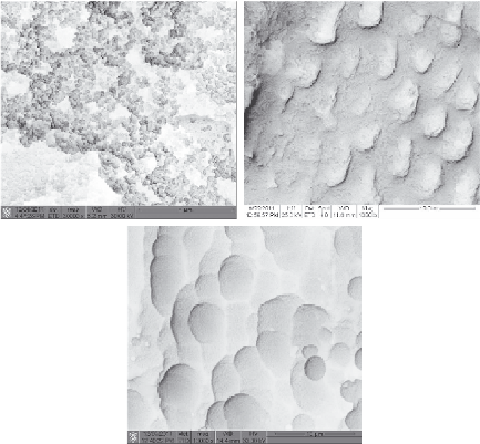
FIGURE 1.1
(a) Nanosized MBG particles, (b) the particles filling dentin tubules, and the induced apatite
mineralization in SBF (c).
time, by controlling the electrospinning conditions they were able to pre-
pare MBG fibers with hollow cores and mesoporous walls; these fibers were
found to be highly bioactive for drug delivery (Hong, Chen, Jing, Fan, Gu, et
al. 2010; Wu, Chang, et al. 2011; Wu and Chang 2012).
It is interesting that MBG could be prepared as uniform spheres with the
size range from nanometer to millimeter. A millimeter-sized MBG sphere
with a well-ordered mesopore channel structure was prepared by using the
method of alginate cross-linking with Ca
2+
ions (see Figure 1.2). The large-
size MBG spheres could not only support the adhesion of bone marrow stro-
mal cells (BMSC) but also control the delivery of proteins (Wu, Zhang, Ke, et
al. 2010). Yun et al. (2009) prepared hierarchically mesoporous-macroporous
MBG spheres with the size of several hundred micrometers in chloroform
by the triblock copolymer templating and sol-gel technique. The spheres
have well interconnected pore structures and excellent
in vitro
bio ac t iv it y.
Mesoporous hollow bioactive glass microspheres with a uniform diameter
Search WWH ::

Custom Search