Biomedical Engineering Reference
In-Depth Information
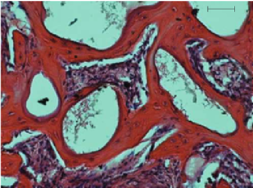
50 µm
FIGURE 1.9
New bone formation around the MBG particles after implanted into the defects of rat femur for
8 weeks (red area: new bone; white area: MBG particles).
(a)
(b)
β-TCP
β-TCP
Akermanite
NB
NB
β-TCP
NB
NB NB
NB
Akermanite
(c)
(d)
NB
NB
NB
NB
NB
β-TCP
NB
Akermanite
NB
β-TCP
β-TCP
NB
Akermanite
FIGURE 2.8
High magnification images of new bone formation and material degradation of (a, c) aker-
manite and (b, c) β-TCP implants after (a, b) 8 and (c, d) 16 weeks (Van Gieson's picrofuchsin
staining of transverse section; NB: new bone). Red color indicates newly formed bone. Original
magnification: 100×.


Search WWH ::

Custom Search