Biomedical Engineering Reference
In-Depth Information
demonstrated that the physiological ions substitution in the crystal struc-
tures of CaP ceramics such as HAp and TCP generates materials with sur-
face bioactivity and superior biological performance comparing traditional
hydroxyapatite and tricalcium phosphate materials (Landi et al. 2008; Xia,
Lindahl, Persson, et al. 2010; Bose et al. 2011; Ballo et al. 2012). Some of these
ions can affect the crystal lattice, and therefore can accelerate the dissolution,
for example, carbonate, silicate, or strontium in HAp. On the other hand,
some additives reduce
in vitro
and
in vivo
the dissolution process, for exam-
ple, fluoride in HAp, magnesium, or zinc in β-TCP (Okazaki et al. 1982; Dhert
et al. 1993; Porter et al. 2004).
Recently, our finding has suggested that the incorporation of silicon
(Si) and strontium (Sr) ions in the HAp biomimetic (Si-HAp and Sr-HAp,
respectively) surfaces improved the surface bioactivity and stimulated bone
apposition in the very early stages of bone healing following implant place-
ment, leading to enhanced osseointegration along the surface of implants
(Figure 5.5) (Ballo et al. 2012). A possible explanation of the higher bioactivity
of Si-HAp and Sr-HAp surfaces is that Si-HAp ceramic has a higher dissolu-
(a)
(b)
IM
20 µm
(c)
IM
1 µm
IM
(d)
IM
100 µm
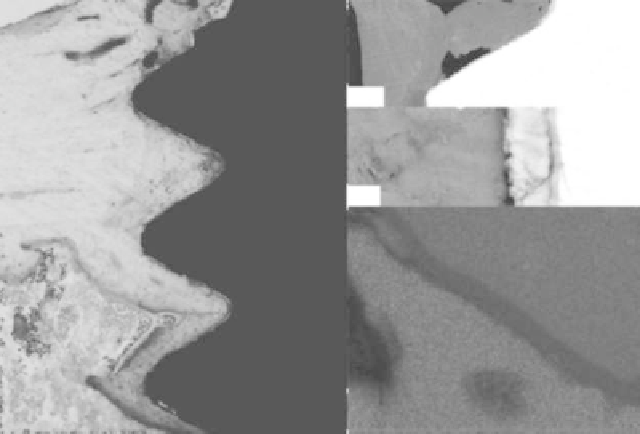
FIGURE 5.5
(
See color insert.
) Sr-HA implants after 28 days of implantation in animal model. (a) Histological
sections revealed mineralized bone growth in the medullary area along and in direct contact
with surfaces was observed (contact osteogenesis). (b, c) Backscatter scanning electron micros-
copy micrographs of the implants after 28 days of healing. (b) Low-magnification image showing
the implant, implant surface, and bone tissue. Osteocyte lacunae and canaliculi were frequently
observed close to the implant surface. (c) Higher-magnification image showing direct contact
was observed. (d) Overlapped element maps of calcium (green), titanium (red), and oxygen
(blue), showing bone formation along the HAp-implant surface at 28 days healing. The enlarged
surface oxide is shown in purple (overlapped blue and red) along the implant perimeter.
Search WWH ::

Custom Search