Biomedical Engineering Reference
In-Depth Information
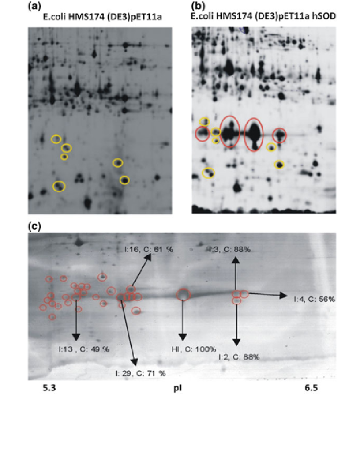
Fig. 1 Spot patterns of samples (150
0
past induction) from a cultivation of E. coli HMS174
(DE3)pET11a (mock strain) (a) and from a cultivation of E. coli HMS174 (DE3)pET11a SOD
(b) on gels separating proteins in the pH range of 3-10 on 18 cm. c Image of a 2D narrow range
gel in the pH range of 5.3-6.5 on 24 cm (same sample as used in b). Red circles indicate spots of
presumable SOD isoforms which were picked for MS identification. Arrows indicate the
identified SOD isoforms
Determination of plasmid-containing cells is done by counting colony-forming
units (cfu) after 24 h of cultivation on LB agar plates containing antibiotics
depending on the resistance marker used for clone screening (100 mg/mL ampi-
cillin or 50 mg/mL kanamycin). The same technique is also used to determine the
genetic stability of integrated cartridges in plasmid-free expression systems.
Search WWH ::

Custom Search