Biomedical Engineering Reference
In-Depth Information
DNA
Lipid
Merged
a)
b)
a/b)
c)
d)
c/d)
e)
f )
e/f )
g)
h)
g/h)
i)
j)
i/j)
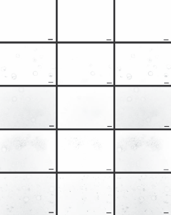
FIGURE 1.6
Fluorescence images of DNA and lipid agent components from representative samples from each of the fol-
lowing groups. (a-b) Mineralized controls, (c-d) plasmid DNA incorporated into PLGA, (e-f) plasmid DNA
coprecipitated with mineral, (g-h) plasmid DNA-lipoplex adsorbed to mineralized films, (i-j) plasmid DNA-
lipoplex coprecipitated with mineral. Distribution of both the plasmid DNA and the lipid transfection agent on
the bone-like mineral was demonstrated by the colocalization of the fluorescent staining in the adsorption and
coprecipitation groups and the absence of staining in the mineralized controls. Scale bars represent 100 um.
(Reprinted from Luong et al.,
Biomaterials,
30(36), 6996-7004, 2009, with permission from Elsevier.)
The method of DNA application affects transfection efficiency. For example, adsorbed
lipoplexes and coprecipitated lipoplexes show significantly different transfection efficien-
cies. Encapsulation of DNA in a calcium phosphate precipitate improves cellular uptake
and produces an enhanced cellular response compared to lipoplexing techniques (Jordan
1996). DNA-lipoplexes coprecipitated with BLM show higher transfection efficiency com-
pared to adsorbed lipoplexes, and coprecipitated naked DNA. This improved transfec-
tion efficiency arises from enhanced cellular uptake and protection from degradation as
a result of cationic lipid complexation, along with the higher availability of apatite at the
surface controlling the rate of release (Luong, McFalls, and Kohn 2009).
Transfection efficiency can also be improved by altering the ionic concentrations of
SBF. For instance, removing Mg ions from the solution improves DNA incorporation and
Search WWH ::

Custom Search