Biomedical Engineering Reference
In-Depth Information
100
: Mirror-polished
: Blasted
80
60
40
20
0
ACP
OAp
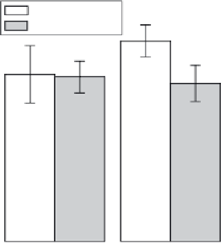
FIGURE 7.27
Bonding strengths of the ACP and OAp coatings fabricated on mirror-polished CP Ti and blasted Ti-6Al-4V
alloy substrates.
by plasma spraying, and (2) the removal of adsorbed gases on the substrate surface in RF
magnetron sputtering and the formation of a functionally graded layer in IBD. Table 7.3
suggests that the bonding strength is affected by heat treatment and immersion in aque-
ous solution such as SBF. The thermal expansion coefficient difference between a calcium
phosphate coating and a Ti substrate together with preferential and partial dissolution
of the coating would decrease the bonding strength. The bonding strength of coatings,
around 60 MPa, is close to that of the epoxy glue used in the pulling tests. As shown in
Figure 7.28,
(65)
the calcium phosphate coating prepared on a Ti substrate by RF magnetron
sputtering remained even after the pulling tests.
(33)
The actual bonding strength of the
coating would be more than 60 MPa. The bonding strength of the thin films to metal sub-
strates should be more precisely evaluated theoretically and experimentally.
The bonding strengths between α-TCP and HAp coatings prepared by thermal CVD and
Ti substrates are 48 and 42 MPa, respectively. These values are slightly smaller than those
of calcium phosphate coatings prepared by sputtering and IBD, but they are higher than
those reported in the coatings prepared by sol-gel and plasma spraying.
Apatite Formation in SBF
Apatite formation on a bioactive glass surface in SBF can be an indicator of
in vivo
bone
formation.
(66,67)
The apatite formation in SBF would be affected by the physical/chemical
properties and roughness of the substrate surface.
CVD can be used to prepare bioceramic coatings such as calcium phosphate and calcium
titanate with a wide variety of surface morphology and crystallographic orientation, and
apatite formation on these coatings in SBF has been reported. Table 7.4 shows the time
required for apatite formation in Hanks' solution over the whole surface of Ca-Ti-O and
Ca-P-O coatings prepared by CVD
(27,28,68)
and other coating methods
(69,70)
on Ti substrates.
Figure 7.29a and b show the surface morphology of the HAp coatings prepared by ther-
mal CVD after immersion in Hanks' solution for 3.6 and 21.6 ks, respectively. Apatite was
identified on the HAp coating after 3.6 ks, and the whole surface of the HAp coating was

Search WWH ::

Custom Search