Biomedical Engineering Reference
In-Depth Information
(a)
(b)
SEI 10.0 kV ×50,000 100 nmWD 8.2 mm
SEI
10.0 kV
×50,000
100 nm
WD 7.9 mm
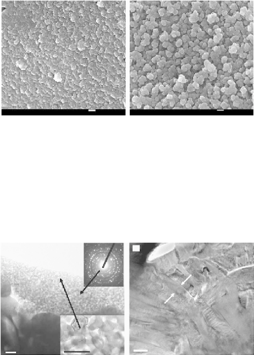
FIGURE 5.22
SEM micrographs of nanosized titania coating on pure titanium prepared by plasma spraying. (a) Low magni-
fication, (b) high magnification. (From Liu et al.,
Biomaterials
, 26, 6143-6150, 2005. With permission.)
is composed of two layers. The outer layer is about 500 nm thick with grains size of less
than 50 nm, whereas the interior layer contains columnar grains with size ranging from
100 to 200 nm, as shown in Figure 5.23b. The SAD pattern discloses a crystalline structure
of the TiO
2
. XRD results further confirm that the coating mainly consists of rutile titania
with a small amount of anatase and TiO
2−
x
suboxide.
Fan et al. have also successfully prepared nanosized titania coatings using vacuum cold
spraying. This system is composed of a vacuum chamber and vacuum pump, an aerosol
room, an accelerating gas feeding unit, a particle-accelerating nozzle, a two-dimensional
work table, and the control unit. TiO
2
particles are accelerated by high-pressure He gas and
(a)
(b)
Outermost layer
Higher magnification
200 nm
100 nm
200 nm
FIGURE 5.23
TEM views of cross section of nanosized titania coating fabricated by plasma spraying and SAD pattern at
selected region. (From Liu et al.,
Biomaterials
, 26, 6143-6150, 2005. With permission.)
Search WWH ::

Custom Search