Biology Reference
In-Depth Information
Figure 5.2
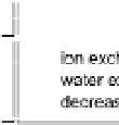
A spider's spinning gland is divided in an A-zone and a
B-zone, where specialized cells secrete silk proteins. Structure
formation and protein assembly are initiated in the spinning
duct, where ion exchange, water extraction, pH decrease and
shear forces are applied (reviewed in Hardy
.
14
and Kenney
et al
et al
.
15
).
Originating from the gland, the spinning dope enters the spinning
duct consisting of three siphon shaped loops. Structure formation
of the proteins already starts in the B-zone of the silk gland, where
�ibrillar structures have been detected re�lecting amyloidogenic
properties such as cross-β structure.
However, the presence of
amyloid-like nano�ibrils in the B-zone has to be treated with caution.
Because the �inal dragline silk belongs to the structural class of
parallel-β silks, the question arises how the structural conversion
takes place and whether these �ibrils play a role in thread formation
or whether they re�lect artefacts of sample preparation.
Within the spinning duct phase separation is induced by
kosmotropic ions leading to removal of water and concentrating the
proteinacous phase. Within the protein phase further folding takes
place, additionally triggered by acidi�ication and shear forces.
16
17-20
Several methods used to investigate structure and assembly of
�ibrous proteins and to analyse amyloidogenic properties of protein
�ibrils can be employed for silk analysis. Studies by atomic force
microscopy or transmission electron microscopy revealed that silk
�ibrils as detected in the B-zone or in
in vitro
assembly reactions,


Search WWH ::

Custom Search