Biology Reference
In-Depth Information
Suspensions of these fibrils form oriented fibres, which give
characteristic “cross-
β
5
(Fig. 4.9). In these
oriented fibres, the long axes of the amyloid-like fibrils seen in the
electron micrographs of Figs. 4.7 and 4.8 are oriented more or less
parallel to the fibre axis.
” X-ray diffraction patterns
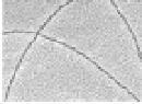
Figure 4.8
Electron micrograph of amyloid-like fibrils derived from a
solution of the cA peptide (experimental conditions as in
Fig. 4.7). Fibrils are rotary shadowed with Pt/Pd at an angle of
7 degrees under high vacuum. Bar =
1000 Å.
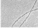
The oriented X-ray pattern (Fig. 4.9) taken from these fibres
indicates the presence of oriented
-sheets in the amyloid-like
fibrils of peptide cA. The presence of reflections corresponding
to periodicities of 4.66 and 10.12 Å indicates the existence of
β
β
-sheets.
20
The strong meridional reflection at 4.66 Å suggests that
the
-strands are perpendicular to
the fibre axis and thus also to the long axis of the amyloid-like fibrils.
The strong equatorial reflection at 10.12 Å, which corresponds to
the intersheet distance, suggests that the packing of the
β
-sheets are oriented so that their
β
-sheets
is parallel to the fibre axis and preferentially oriented. This X-ray
pattern closely resembles typical cross-
β
β
patterns
21
taken from
amyloid fibres (ref. 22, and references therein).


Search WWH ::

Custom Search