Biology Reference
In-Depth Information
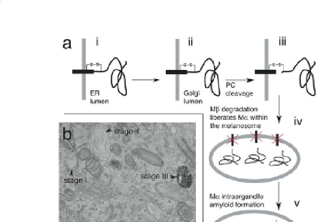
Figure 9.1
Pmel17 trafficking and function in melanin biosynthesis
and melanosome biogenesis. (a) Pmel17 is synthesized as
a transmembrane protein in the ER (i), it is trafficked first
to the Golgi (ii), and finally to the melanosome. Proprotein
convertase (PC) cleavage in a post-Golgi compartment creates
a lumenal fragment, M
α
, and a transmembrane fragment, Mβ.
M
α
remains disulphide-bonded to Mβ to prohibit aberrant M
α
amyloidogenesis (iii). Degradation of Mβ releases M
α
from the
α
membrane (iv), enabling M
to form amyloid fibres within
the melanosome organelle (v). M
α
amyloid fibres orchestrate
the synthesis of melanin from tyrosine-derived reactive
indolequinones and protect the melanocyte from melanin-
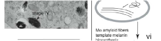
associated toxicity (vi). (b) Electron micrograph depicting
the four-stage process of melanosome maturation. Stage I:
melanosomes are vesicles of endosomal origin. Stage II: M
α
amyloid fibres form within the nascent melanosome. Stage III:
melanin appears along M
α
fibres. Stage IV: melanin occludes
the M
fibre structure in melanosomes. Part (a) and figure
legend reproduced with permission from Elsevier.
α
5
Electron
micrograph was generated by Ilse Hurbain and Graça Raposo.

Search WWH ::

Custom Search